. 2013;8(1):e52384.
doi: 10.1371/journal.pone.0052384. Epub 2013 Jan 9.

Activated ERM protein plays a critical role in drug resistance of MOLT4 cells induced by CCL25

Affiliations

Activated ERM protein plays a critical role in drug resistance of MOLT4 cells induced by CCL25

Li Zhang et al. PLoS One. 2013.

Abstract

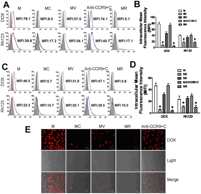
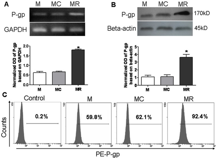
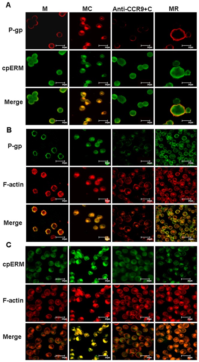
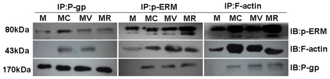
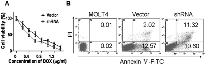
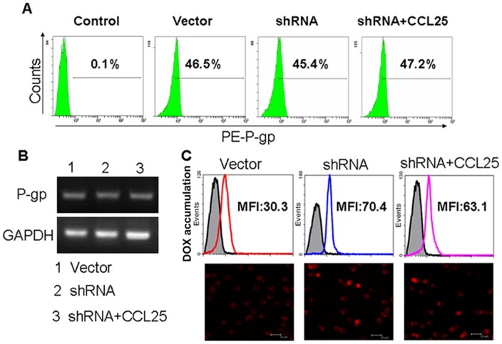
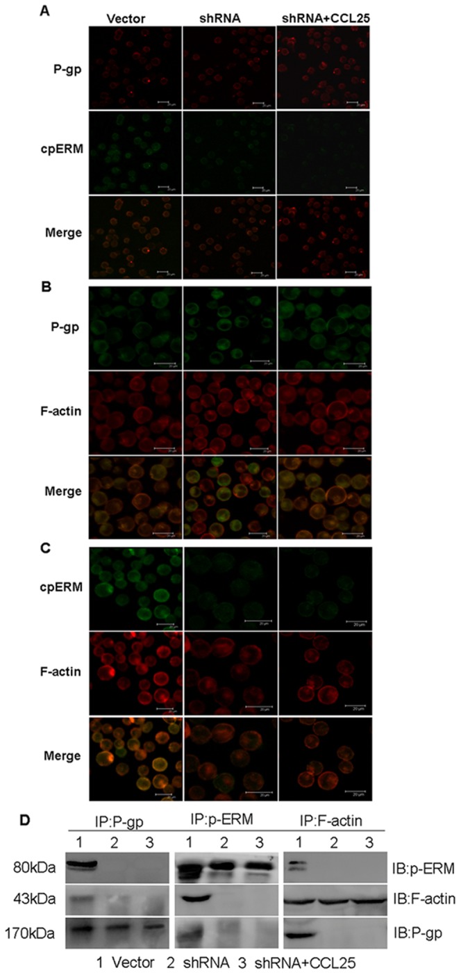
We have previously demonstrated that the CCR9/CCL25 signaling pathway plays an important role in drug resistance in human acute T-lymphocytic leukemia (T-ALL) by inducing activation of ERM protein with polarized distribution in T-ALL cell line MOLT4. However, the mechanism of action of the activated ERM protein in the drug resistance of MOLT4 cells induced by CCL25 remains uncharacterized. Here we investigated the mechanism of CCR9/CCL25-initiated drug resistance in CCR9-high-expressing T-ALL cells. Our results showed that 1) the function of P-gp was increased after treatment with CCL25; 2) P-gp colocalized and co-immunoprecipitated with p-ERM and F-actin in CCL25 treated cells; and 3) ERM-shRNA conferred drug sensitivity coincident with release of ERM interactions with P-gp and F-actin after treatment with CCL25. These data suggest it is pivotal that P-gp associate with the F-actin cytoskeleton through p-ERM in CCR9/CCL25 induced multidrug resistance of T-ALL cells. Strategies aimed at inhibiting P-gp-F-actin cytoskeleton association may be helpful in increasing the efficiency of therapies in T-ALL.


Conflict of interest statement

Competing Interests: The authors have declared that no competing interests exist.

Figures

Figure 1
Figure 1. P-gp functions in MOLT4 cells treated with different factors.
(A) Intracellular drug accumulation in untreated MOLT4 cells or MOLT4 cells treated with CCL25/verapamil/anti-CCR9 by FCM. These cells were incubated with DOX or Rh123, washed and the MFI determined by flow cytometry using MR cell line as a control. (B) The accumulation of DOX and Rh123 in MOLT4 cells. Values are mean ± SD. *, P<0.05. (C) Efflux assessment of DOX and Rh123 in MOLT4 cells detected by FCM. Cells were incubated with DOX or Rh123 as described above followed by washing. After 1 h of dye efflux at 37°C in the medium without DOX or Rh123, cells were washed twice with cold PBS and immediately analyzed using the same procedure as for the accumulation experiments. (D) The efflux of DOX and Rho123 in MOLT4 cells. Values are mean ± SD. *, p<0.05. (E) Intracellular accumulations in MOLT4 cells detected by LSCM. Cells were washed with PBS, incubated with or without verapamil before treatment with DOX, and then the cells were treated with CCL25 or anti-CCR9 antibody and CCL25. Confocal images (top); Nomarski differential interference contrast images (middle); and overlays of confocal and differential interference contrast images (bottom). Bar = 20 µm. All the results presented are the means of three independent experiments. M, MOLT4; MC, MOLT4 cells treated with CCL25; MV, MOLT4 cells treated with CCL25 and VRP; MR, MOLT4 cells treated with DOX for 9 months.
Figure 2
Figure 2. P-gp expression induced by CCL25 in MOLT4 cells.
(A) RT-PCR and (B) Western blot analyses for P-gp expression induced by CCL25 in MOLT4 cells. MR cells were used as positive control. Amplification of GAPDH was used as control for RT-PCR assays. For loading control in western blot, membranes were incubated with monoclonal anti-β-actin antibody. (C). Expression of P-gp in cells stained with PE-anti-human MDR1 antibody by FCM. Values presented are the means of four independent experiments.
Figure 3
Figure 3. Colocalization of P-gp, p-ERM and F-actin in MOLT4 cells treated with CCL25 by confocal.
Shown are the subcellular localizations of (A) P-gp (red) and p-ERM (green), (B) P-gp (green) and F-actin (red), and (C) p-ERM (green) and F-actin (red) in MOLT4 cells treated with CCL25 or anti-CCR9 and CCL25 together. All the images showed that P-gp and p-ERM or F-actin colocalized in CCL25 treated cells, and this colocalization could be abolished by anti-CCR9 antibody. Bar = 20 µm. The results represented the means of four independent experiments.
Figure 4
Figure 4. P-gp, ERM and F-actin co-immunoprecipitation induced by CCL25 in MOLT4 cells.
Western blot for p-ERM, F-actin, and P-gp in P-gp (left panel), p-ERM (middle panel) and F-actin (right panel) immunoprecipitations, respectively, from lysates of MOLT4 cells in the presence or absence of CCL25.
Figure 5
Figure 5. Effects of ERM-shRNA on ERM expression.
MOLT4 cells were transfected with ERM-shRNAs and cells were selected in G418 (200 µg/ml) for 2 weeks. After 6–8 weeks with G418 selection, the clones were selected for identification by RT-PCR and Western blot.
Figure 6
Figure 6. Cell viability and apoptosis analysis in ERM-silenced MOLT4 cells.
(A) Cell viability was lower in ERM-silenced cells compared to empty vector-transfected cells. (B) The apoptosis rate in shRNA transfected cells was increased compared with empty vector-transfected cells.
Figure 7
Figure 7. Expression and functionality of P-gp in ERM-silenced MOLT4 cells treated with CCL25.
(A) Expression of P-gp by FCM. (B) Expression of P-gp by RT-PCR. (C) DOX accumulation by FCM (upper) and LSCM (bottom). Bar = 20 µm.
Figure 8
Figure 8. Role of P-gp-p-ERM-F-actin association in ERM-silenced MOLT4 cells treated by CCL25.
Shown are the subcellular localizations of (A) P-gp (red) and p-ERM (green), (B) P-gp (green) and F-actin (red), (C) p-ERM (green) and F-actin (red) in ERM-silenced MOLT4 cells treated by CCL25. All images showed P-gp and p-ERM or F-actin distribution on the cells. Bar = 20 µm. (D) Co-immunoprecipitation analysis of ERM-silenced MOLT4 cells, either untreated or pretreated with CCL25.

Similar articles

Cited by

References

    1. Pui CH, Behm FG, Crist WM (1993) Clinical and biologic relevance of immunologic marker studies in childhood acute lymphoblastic leukemia. Blood 82: 343–362. - PubMed
    1. Willemse MJ, Seriu T, Hettinger K, d'Aniello E, Hop WC, et al. (2002) Detection of minimal residual disease identifies differences in treatment response between T-ALL and precursor B-ALL. Blood 99 (12) 4386–4393. - PubMed
    1. Gottesman MM, Ling V (2006) The molecular basis of multidrug resistance in cancer: the early years of P-glycoprotein research. FEBS Lett 580: 998–1009. - PubMed
    1. Sharon FJ (2008) ABC multidrug transporters: structure, function and role in chemoresistance. Pharmacogenomics 9 (1) 105–127. - PubMed
    1. Meschini S, Calcabrini A, Monti E, Del Bufalo D, Stringaro A, et al. (2000) Intracellular P-glycoprotein expression is associated with the intrinsic multidrug resistance phenotype in human colon adenocarcinoma cells. Int J Cancer 87 (5) 615–628. - PubMed

Publication types

MeSH terms

LinkOut - more resources