. 2013;8(1):e52543.
doi: 10.1371/journal.pone.0052543. Epub 2013 Jan 9.

Sickle erythrocytes target cytotoxics to hypoxic tumor microvessels and potentiate a tumoricidal response

Affiliations

Sickle erythrocytes target cytotoxics to hypoxic tumor microvessels and potentiate a tumoricidal response

David S Terman et al. PLoS One. 2013.

Abstract

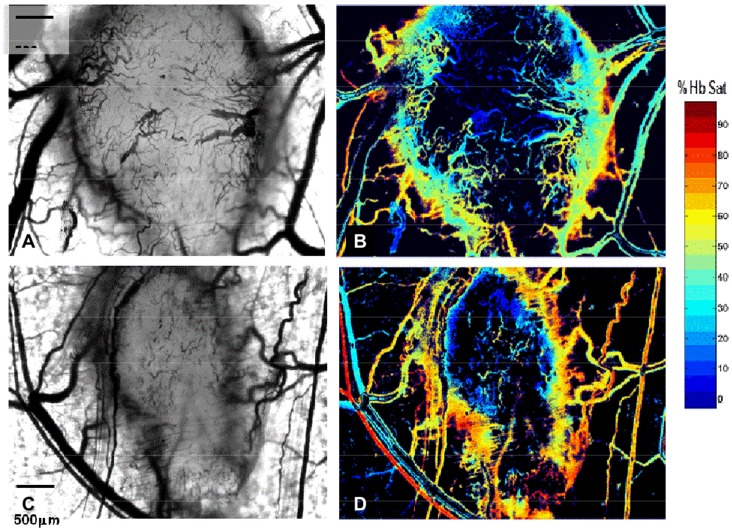
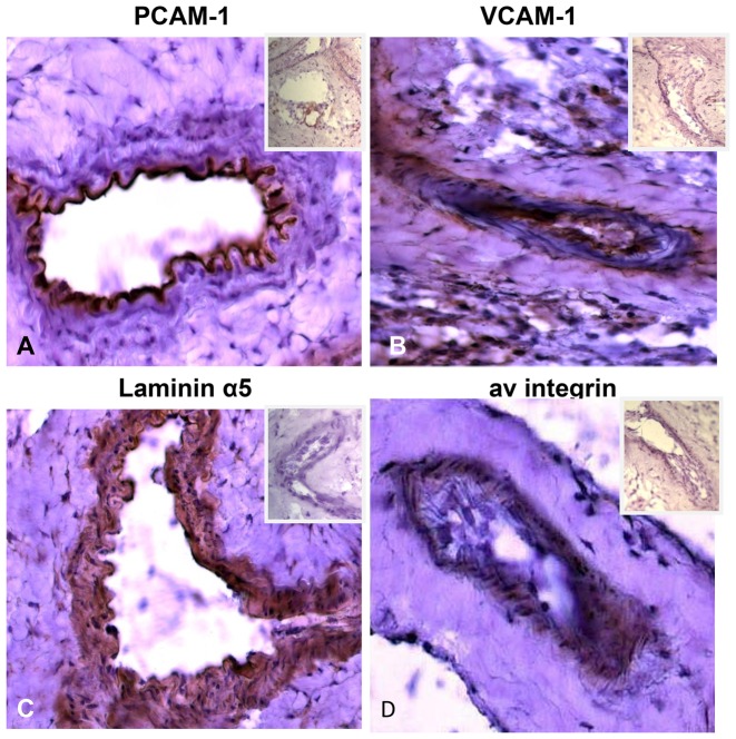
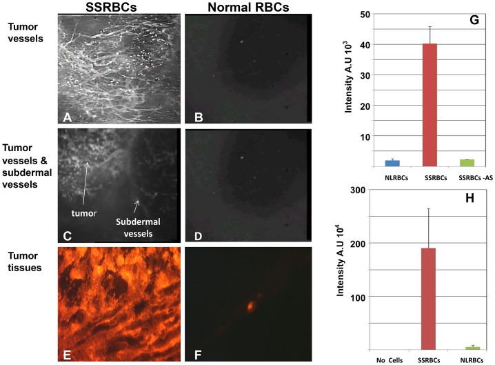
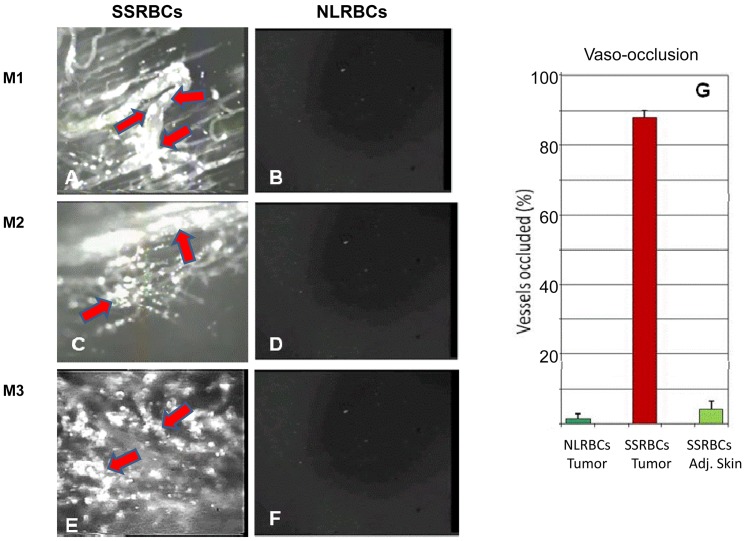
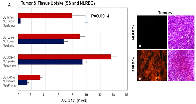
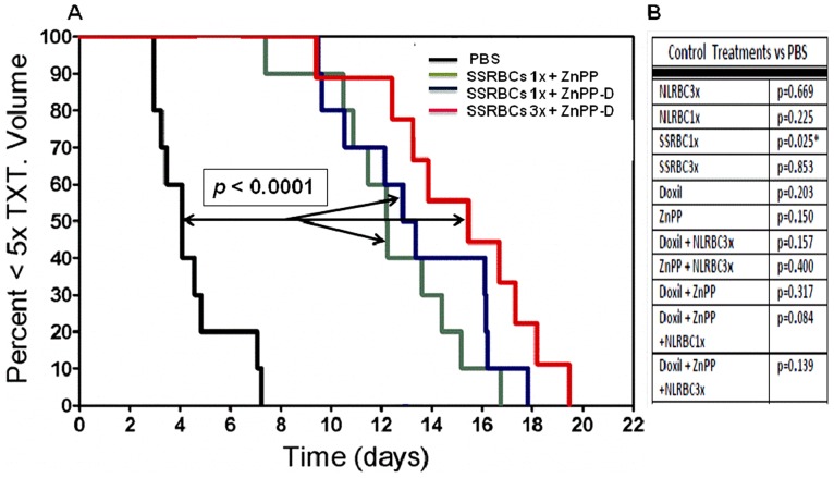
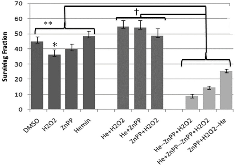
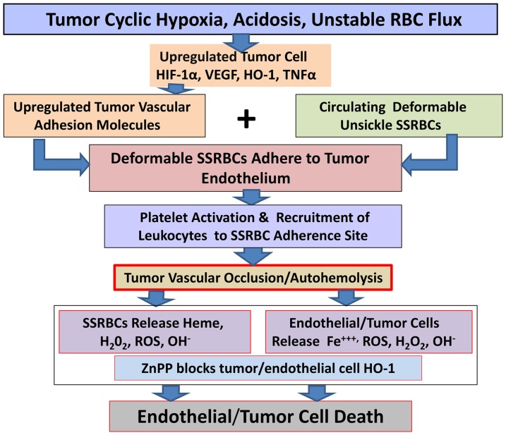
Resistance of hypoxic solid tumor niches to chemotherapy and radiotherapy remains a major scientific challenge that calls for conceptually new approaches. Here we exploit a hitherto unrecognized ability of sickled erythrocytes (SSRBCs) but not normal RBCs (NLRBCs) to selectively target hypoxic tumor vascular microenviroment and induce diffuse vaso-occlusion. Within minutes after injection SSRBCs, but not NLRBCs, home and adhere to hypoxic 4T1 tumor vasculature with hemoglobin saturation levels at or below 10% that are distributed over 70% of the tumor space. The bound SSRBCs thereupon form microaggregates that obstruct/occlude up to 88% of tumor microvessels. Importantly, SSRBCs, but not normal RBCs, combined with exogenous prooxidant zinc protoporphyrin (ZnPP) induce a potent tumoricidal response via a mutual potentiating mechanism. In a clonogenic tumor cell survival assay, SSRBC surrogate hemin, along with H(2)O(2) and ZnPP demonstrate a similar mutual potentiation and tumoricidal effect. In contrast to existing treatments directed only to the hypoxic tumor cell, the present approach targets the hypoxic tumor vascular environment and induces injury to both tumor microvessels and tumor cells using intrinsic SSRBC-derived oxidants and locally generated ROS. Thus, the SSRBC appears to be a potent new tool for treatment of hypoxic solid tumors, which are notable for their resistance to existing cancer treatments.


Conflict of interest statement

Competing Interests: The authors have read the journal's policy and have the following conflicts: DST is sole inventor of US patent 7,803,637 filed August 30, 2000, issued September 28, 2010. All other authors have no competing interests. Dr. David S. Terman is employed by Jenomic. This does not alter his adherence to all the PLOS ONE policies on sharing data and materials. Reovirus Dearing Type 3 was provided by Oncolytics Biotech Inc. (Calgary, Canada). This does not alter the authors' adherence to all the PLOS ONE policies on sharing data and materials.

Figures

Figure 1
Figure 1. Eight day old 4T1 carcinoma is vascularized and hypoxic.
Intravital microscopy of two eight day old 4T1 tumors implanted in the dorsal skin window chamber viewed with light microscopy shows diffuse tumor microvascularity (panels A, C). Corresponding hyperspectral imaging of the same tumors exhibits hemoglobin saturations ≤10% over a 70% of the tumor surfaces (B,D). Magnification 5×.
Figure 2
Figure 2. Expression of adhesion molecules on 4T1 tumor vascular endothelium.
Frozen sections of 4T1 tumors stained with antibodies against various adhesion molecules shows significant endothelial expression of PECAM-1 (A), ICAM-4 (B), laminin α5 (C). αv integrin (D). Secondary antibodies alone used as negative controls to stain the same tumor sections are shown in the inset of each panel. Magnification 40×.
Figure 3
Figure 3. SSRBCs but not NLRBCs accumulate in tumor microvessels within 30 minutes after injection.
Intravital microscopy of the vasculature of 8-day old 4T1 tumors implanted in the dorsal skin window chamber within 30 minutes after infusion of mice with SSRBCs (A, C, E) or NLRBCs (B,D,F) shows the accumulation of SSRBCs but not NLRBCs in the tumor blood vessels and tumor parenchyma (A,B,E,F). At the same time, SSRBC uptake is observed in the tumor vessels, there is minimal uptake in the adjacent subdermal blood vessels (C). There is also minimal uptake of NLRBCs in adjacent subdermal blood vessels (D) (Magnification 5×). Thirty minutes after infusion, the uptake of fluorescently-labeled SSRBCs (n = 5) or NLRBCs (n = 5) in tumor vessels (G) and tumor parenchyma (H) is quantitated in still video images (fluorescence intensity (FI) at Magnification 20×). SSRBCs (n = 6) show significantly greater mean FI in tumor vessels and parenchyma (G and H respectively) compared to subdermal skin vessels or NLRBCs (n = 3) (p = 0.00001 for FI of SSRBCs in tumor vessels and tumor parenchyma vs. respective controls in both G and H). Abbreviations in legend: AS: adjacent subdermal skin vessels.
Figure 4
Figure 4. SSRBCs but not NLRBCs form microaggregates and occlude tumor microvessels.
Thirty minutes after infusion of SSRBCs or NLRBCs into mice bearing eight day old 4T1 tumors, diffuse tumor vaso-occlusion is evident in mice injected with SSRBCs (A,C,E) but not NLRBCs (B,D,F). (Magnification 10× was used in panels 1–4 and 20× in panels 5 and 6). Arrows indicate SSRBC adhesion to vascular walls, microaggregate formation and partial or complete microvessel occlusion. The mice injected with SSRBCs (n = 5) showed a significantly greater percentage of occluded tumor vessels compared to NLRBCs (n = 5) or adjacent subdermal skin vessels (G) (p = 0.00001 for SSRBCs in tumor vessels vs. NLRBCs or adjacent subdermal skin vessels). Magnification 20× was used for quantitation of tumor microvessel occlusion. Abbreviations in legend: Adj. skin: adjacent subdermal skin vessels.
Figure 5
Figure 5. SSRBCs accumulate to a significantly greater degree in tumors compared to NLRBCs.
RFP-labeled SSRBCs (n = 4) or NLRBCs (n = 2) were injected into mice bearing eight day old 4T1 tumors. Twenty four hours later tumors and organs were collected and RFP fluorescence quantitated on sections of tumors and organs. The uptake of SSRBCs in tumors is significantly greater than NLRBCs (p = 0.0014) (A, B, D). In contrast, the uptake of SSRBCs and NLRBCs is not significantly different in the spleen, lungs and kidneys (p>0.05) (A) (Magnification 5×). H&E tumor sections from SSRBC-treated mouse show focal areas of cytoplasmic eosinophilia consistent with ischemia (E) not present in tumors treated with NLRBCs (C) (Magnification 20×). Abbreviations in legend: negTumor, negLung, negSpleen, negKidney mean mice injected with NLRBCs or SSRBCs without RFP label.
Figure 6
Figure 6. SSRBCs but not NLRBCs combined with prooxidants ZnPP and ZnPP-D induce a tumoricidal response in 4T1 bearing mice.
The fraction of mice with tumors <5× the pretreatment volumes versus time is shown (n = 10 for each treatment group). All three groups treated with SSRBCs combined with ZnPP or ZnPP-D show significant tumor growth delay compared to PBS controls. In the adjacent Table, a control experiment shows that tumor bearing animals receiving SSRBCs 1× or 3× alone, NLRBC 1× or 3× alone, NLRBCs 1× or 3× with ZnPP or ZnPP-D, ZnPP alone or Doxil + ZnPP exhibited no significant tumor growth delay versus the PBS control. * indicates that mice receiving SSRBC1× alone displayed significantly accelerated tumor growth compared to the PBS control.
Figure 7
Figure 7. Tumoricidal effect of the combination of hemin, H2O2 and ZnPP in a clonogenic tumor survival model.
Three agent regimens consisting of pre-treating 4T1 cells with i) hemin alone or combined with ZnPP for 2 hrs followed by the combination of ZnPP and H2O2 for 2 hrs or, ii) ZnPP for 2 hours followed by the combination of hemin and H2O2 for 2 hours induced significant tumor cell death compared to each agent individually (**p<0.0002) and any two of these agents used simultaneously (†p<0.0001). Clonogenic survival is shown as a mean of three independent experiments with standard error (SE) indicated. See Table S2 for protocol used in these studies.
Figure 8
Figure 8. Schematic depiction of proposed pathophysiology of tumor killing induced by SSRBCs and the HO-1 inhibitor ZnPP.
The hypoxic and acidic tumor milieu activates HIF1α, which, in turn, stimulates VEGF and HO-1 expression and the production of TNFα. TNFα upregulates several adhesion molecules on tumor endothelium, including several endothelial cognate adhesion ligands for the major adhesion receptors expressed on SSRBCs. Deformable non-sickled SS RBCs adhere to the activated endothelium of the tumor vasculature, along with leukocytes to form microaggregates leading to tumor vascular obstruction/occlusion. Entrapped SSRBCs release SS hemoglobin which is converted rapidly to methemoglobin and cleaved to liberate free heme. Hydrophobic and lipophilic heme and/or heme-nitrosyl complexes permeate tumor and endothelial cell membranes where they catalytically oxidize lipids, proteins and DNA causing cell death. In the presence of ZnPP, a competitive inhibitor of HO-1, intracellular heme and oxidative products such as reactive oxygen and nitrogen species (ROS and RNS) are free to exert their potent oxidative function leading to tumor and endothelial cell death.

Similar articles

Cited by

References

    1. Kerbel RS (2008) Tumor angiogenesis. N Engl J Med 358: 2039–2049. - PMC - PubMed
    1. Dewhirst MW (2009) Relationships between cycling hypoxia, HIF-1, angiogenesis and oxidative stress. Radiat Res 172: 653–665. - PMC - PubMed
    1. Bristow RG, Hill RP (2008) Hypoxia, DNA repair and genetic instability. Nat Rev Cancer 8: 180–192. - PubMed
    1. Pries AR, Hopfner M, Noble F, Dewhirst MW, Secomb TW (2010) The shunt problem: control of functional shunting in normal and tumour vasculature. Nat Rev Cancer 10: 587–593. - PMC - PubMed
    1. Wilson WR, Hay MP (2011) Targeting hypoxia in cancer therapy. Nat Rev Cancer 11: 393–410. - PubMed

Publication types

MeSH terms