. 2013;8(1):e52980.
doi: 10.1371/journal.pone.0052980. Epub 2013 Jan 10.

Nuclear legumain activity in colorectal cancer

Affiliations

Nuclear legumain activity in colorectal cancer

Mads H Haugen et al. PLoS One. 2013.

Erratum in

  • PLoS One. 2013;8(9). doi:10.1371/annotation/05c95441-890f-4707-a1bc-c4d386561191

Abstract

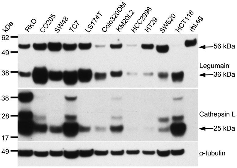
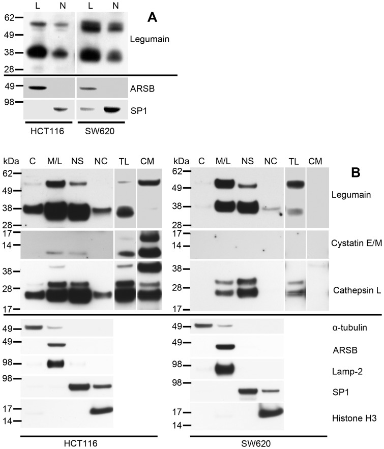
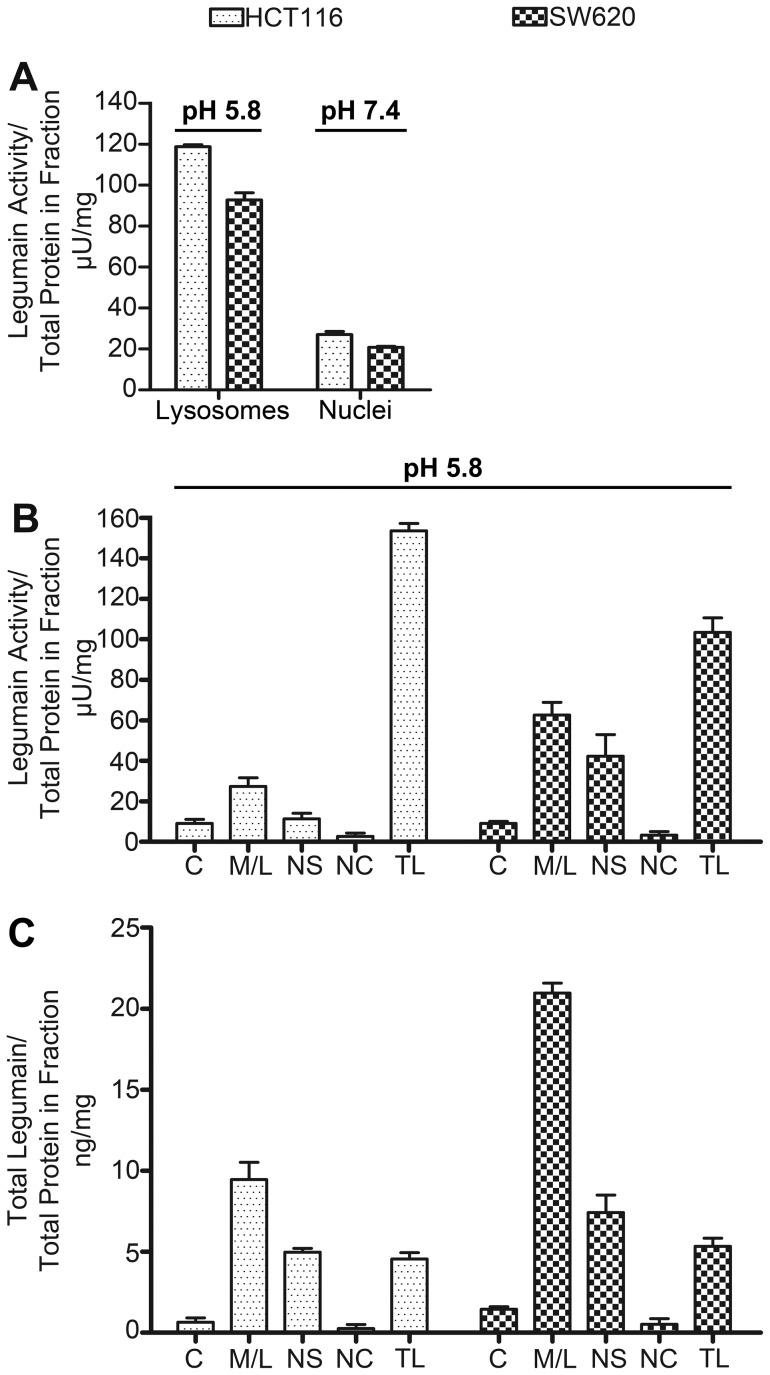
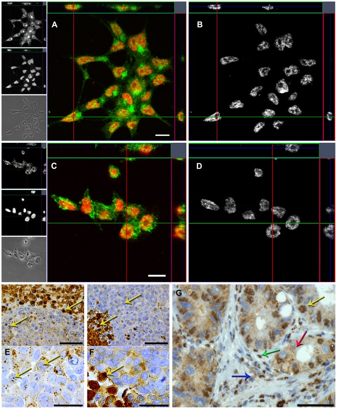
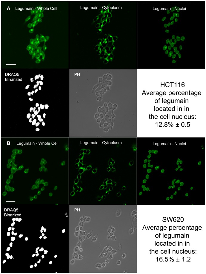
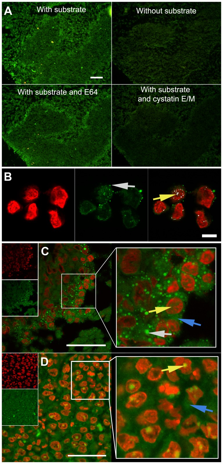
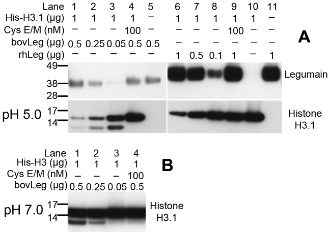
The cysteine protease legumain is involved in several biological and pathological processes, and the protease has been found over-expressed and associated with an invasive and metastatic phenotype in a number of solid tumors. Consequently, legumain has been proposed as a prognostic marker for certain cancers, and a potential therapeutic target. Nevertheless, details on how legumain advances malignant progression along with regulation of its proteolytic activity are unclear. In the present work, legumain expression was examined in colorectal cancer cell lines. Substantial differences in amounts of pro- and active legumain forms, along with distinct intracellular distribution patterns, were observed in HCT116 and SW620 cells and corresponding subcutaneous xenografts. Legumain is thought to be located and processed towards its active form primarily in the endo-lysosomes; however, the subcellular distribution remains largely unexplored. By analyzing subcellular fractions, a proteolytically active form of legumain was found in the nucleus of both cell lines, in addition to the canonical endo-lysosomal residency. In situ analyses of legumain expression and activity confirmed the endo-lysosomal and nuclear localizations in cultured cells and, importantly, also in sections from xenografts and biopsies from colorectal cancer patients. In the HCT116 and SW620 cell lines nuclear legumain was found to make up approximately 13% and 17% of the total legumain, respectively. In similarity with previous studies on nuclear variants of related cysteine proteases, legumain was shown to process histone H3.1. The discovery of nuclear localized legumain launches an entirely novel arena of legumain biology and functions in cancer.


Conflict of interest statement

Competing Interests: The authors have declared that no competing interests exist.

Figures

Figure 1
Figure 1. Expression of legumain and cathepsin L in CRC cell lines.
Immunoblots of cell lysates from a panel of CRC cell lines demonstrated high variability in the total amount of legumain and cathepsin L, and also in the presence of the different mature forms. HCT116 and SW620 cells were particularly interesting as they show mutually exclusive high amount of the active (36 kDa) and inactive pro-form (56 kDa) of legumain, respectively. Uncut immunoblots (Fig. S2A).
Figure 2
Figure 2. Legumain, cystatin E/M and cathepsin L expressions in subcellular fractions of HCT116 and SW620 cells.
(A) Immunoblots of legumain in lysosomal (L) and nuclear (N) fractions enriched from HCT116 and SW620 cells using density gradient centrifugation. All lanes were loaded with 15 µg total protein from each fraction. Purity controls of the subcellular fractions were assessed by staining for ARSB (soluble lysosomal protein) and SP1 (nuclear transcription factor). (B) Immunoblots of legumain (top panels), cystatin E/M (second panels) and cathepsin L (third panels) in enriched subcellular compartments isolated from HCT116 and SW620 cells using a commercial kit: Cytosol (C), membranes/lysosomes (M/L), nuclear soluble (NS), nuclear chromatin bound (NC), total lysate (TL) and conditioned media (CM). All lanes were loaded with 15 µg total protein from each fraction, except conditioned media where proteins precipitated from 1 ml was loaded. Purity controls of the different subcellular fractions were assessed by staining for α-tubulin (cytosolic protein), ARSB (soluble lysosomal protein), lamp-2 (lysosome membrane-associated protein), SP1 (nuclear transcription factor) and histone H3 (nuclear chromatin bound protein). Uncut immunoblots of legumain and cathepsin L (Fig. S2C).
Figure 3
Figure 3. Proteolytic activity and quantity of legumain in subcellular fractions of HCT116 and SW620 cells.
(A) Proteolytic activity of legumain determined by substrate cleavage (Z-Ala-Ala-Asn-NHMec) relative to total protein content of each subcellular compartment of HCT116 (dotted bars) and SW620 (chequered bars) cells after density gradient centrifugations. Lysosomal and nuclear fractions were prepared and analyzed at pH 5.8 and 7.4, respectively, demonstrating proteolytic activity of legumain in both lysosomal and nuclear fractions of both cell lines at both pH conditions, although highest in the lysosomal compartment assayed at pH 5.8. (B) Proteolytic activity of legumain measured at pH 5.8 in subcellular fractions prepared by a commercial kit was found to be highest in the M/L fractions, but was also clearly present in the NS fractions and observed with only minor activity in the C fractions of both cell lines. Extracellular legumain did not demonstrate any activity in either cell line (data not shown). (C) Total legumain amounts (pro- and active form) measured by ELISA in subcellular fractions (isolated using a commercial kit) and calculated relative to the total protein content in each fraction were also highest in the M/L fractions, yet clearly present in the NS fraction.
Figure 4
Figure 4. Subcellular localization of legumain in HCT116 and SW620 cells, subcutaneous xenografts, and human CRC tumor tissue.
(A to D) Immunofluorescence staining of legumain (green and top left panels) and nuclei (red and middle left panels) in HCT116 (A) and SW620 (C) cells cultured on glass slides and visualized as orthogonal slices of z-stacks by confocal laser scanning microscopy. By using image arithmetics with the binarized capture of corresponding DRAQ5™-positive nuclei as a mask, nuclear legumain representative signals were extracted from all optical sections and visualized in grayscale as orthogonal slices for HCT116 (B) and SW620 (D). Scale bars represent 10 µm. Specificity of immunofluorescence signals was verified by incubation with secondary antibodies only, yielding no signal (Fig. S3A and S3B). (E and F) When grown as subcutaneous xenografts in mice, immunohistochemical staining of legumain in HCT116 (E) cells demonstrated a much more granulated staining pattern than in SW620 (F) cells. However, both cell lines exhibited areas of strong legumain expression and also in the nuclei (yellow arrows). Scale bars represent 50 (top panels) and 25 (bottom panels) µm. H/E stain (Fig. S3C and S3D). Goat-IgG isotype control showed low staining (Fig. S3E and S3F). (G) Immunohistochemical staining of legumain in paraffin-embedded section from a representative CRC tumor biopsy showing nuclear staining of legumain in some, but not all, epithelial cells (i.e. carcinoma cells; yellow arrow) and stromal cells (green arrow). Epithelial cells also exhibited marked granulated staining in the cytoplasm (red arrow), whereas stromal cells showed much weaker staining outside the nucleus (blue arrow). Scale bar represent 50 µm. Goat-IgG isotype control showed no staining (Fig. S3G).
Figure 5
Figure 5. Percentage of expressed legumain located in the nucleus.
(A and B) Representative pictures of HCT116 (A) and SW620 (B) cells by one optical slice from one of five independent z-stacks each containing 30–40 immunofluorescently labeled cells using legumain-specific antibodies (green), with DRAQ5™ counter-stained nuclei (binarized; white). By means of a semi-automated procedure in Image J the captures of nuclei (binarized) was used as a mask to separate the nuclear (top right) from the cytoplasmic (top middle) signal components of the total (top left) signal representing expression of legumain. The total signal from legumain fluorescence in each optical slice was summarized from all five z-stacks enabling for the estimation of the expressed amount of legumain in the nuclear compartment. Statistical errors in the calculations are reported as standard error of the mean of the five independent z-stacks. Scale bar represents 20 µm.
Figure 6
Figure 6. In situ legumain activity in cultured cells and subcutaneous xenografts.
(A) In situ proteolytic activity (green) captured by fluorescence microscopy imaging of adjacent cryosections from a HCT116 subcutaneous xenograft incubated with (top left) and without (top right) legumain substrate, legumain substrate and E64 (lower left) or legumain substrate and recombinant cystatin E/M (lower right), demonstrating the specificity of the synthetic peptide Suc-Ala-Ala-Asn-NHNapOME utilized as legumain substrate. All pictures were taken using true colors, after the same incubation time and with identical microscope and camera settings. Scale bar represents 100 µm. Subcutaneous xenografts with SW620 cells (Fig. S3J). (B) Subcellular localization of active legumain (green) in HCT116 cells (made from cryosections after mounting in OCT-medium) with nuclei stained by DAPI (red) and analyzed by confocal laser scanning microscopy. This showed granulated activity inside (yellow arrow) and outside (gray arrow) of the nucleus. Localization in the nucleus was confirmed by co-localization (white) of legumain activity and the nuclear counter-stain (right panel). Scale bar represents 10 µm. HCT116 cells incubated without substrate, or with substrate and cystatin E/M, showed no signals (Fig. S3H and I, respectively). (C and D) Legumain activity (green) in cryosections from subcutaneous xenografts with nuclei stained with DAPI (red) and analyzed by confocal laser scanning microscopy. Subcutaneous xenograft from HCT116 cells (C) showed similar results as in cultured cells with intense granulated activity (gray arrow) although less distinct activity in the cytoplasm (blue arrow) and within the nucleus (yellow arrow) was also observed. However, in the subcutaneous xenograft of SW620 cells (D) majorly diffuse legumain activity was observed in the cell cytoplasm (blue arrow), while in the nucleus this was more concentrated (yellow arrow). Scale bars represent 50 µm.
Figure 7
Figure 7. Cleavage of histone H3.1 by active legumain.
(A) Immunoblots showing the cleavage of intact (lane 10) recombinant human histone H3.1 in a dose dependent manner by purified mature 36 kDa bovine legumain (bovLeg, lane 1–3) and auto-activated intermediate form (46 kDa) of recombinant human legumain (rhLeg, lane 6–8). The addition of recombinant human cystatin E/M (lane 4 and 8) efficiently blocked legumain activity and resulted in almost complete rescue of histone H3.1 from proteolytic cleavage. Uncut immunoblots (Fig. S2D). (B) Immunoblot of histone H3.1 showing the dose-dependent production of a 12 kDa cleavage product after incubation of recombinant histone H3.1 with fully mature 36 kDa bovine legumain in a buffer with pH 7.0 (lane 1–3). Addition of recombinant human cystatin E/M efficiently blocked legumain activity and resulted in virtually no formation of the 12 kDa cleavage product (lane 4).

Similar articles

Cited by

References

    1. Rawlings ND, Barrett AJ, Bateman A (2010) MEROPS: the peptidase database. Nucleic Acids Res 38: D227–233. - PMC - PubMed
    1. Kembhavi AA, Buttle DJ, Knight CG, Barrett AJ (1993) The Two Cysteine Endopeptidases of Legume Seeds: Purification and Characterization by Use of Specific Fluorometric Assays. Arch Biochem Biophys 303: 208–213. - PubMed
    1. Hara-Nishimura I, Takeuchi Y, Nishimura M (1993) Molecular Characterization of a Vacuolar Processing Enzyme Related to a Putative Cysteine Proteinase of Schistosoma mansoni. Plant Cell 5: 1651–1659. - PMC - PubMed
    1. Chen JM, Dando PM, Rawlings ND, Brown MA, Young NE, et al. (1997) Cloning, isolation, and characterization of mammalian legumain, an asparaginyl endopeptidase. J Biol Chem 272: 8090–8098. - PubMed
    1. Dall E, Brandstetter H (2012) Activation of legumain involves proteolytic and conformational events, resulting in a context- and substrate-dependent activity profile. Acta Crystallogr F 68: 24–31. - PMC - PubMed

Publication types

MeSH terms

Substances