

Purification of mitochondrial proteins HSP60 and ATP synthase from ascidian eggs: implications for antibody specificity
- PMID: 23326373
- PMCID: PMC3542361
- DOI: 10.1371/journal.pone.0052996
Purification of mitochondrial proteins HSP60 and ATP synthase from ascidian eggs: implications for antibody specificity
Abstract
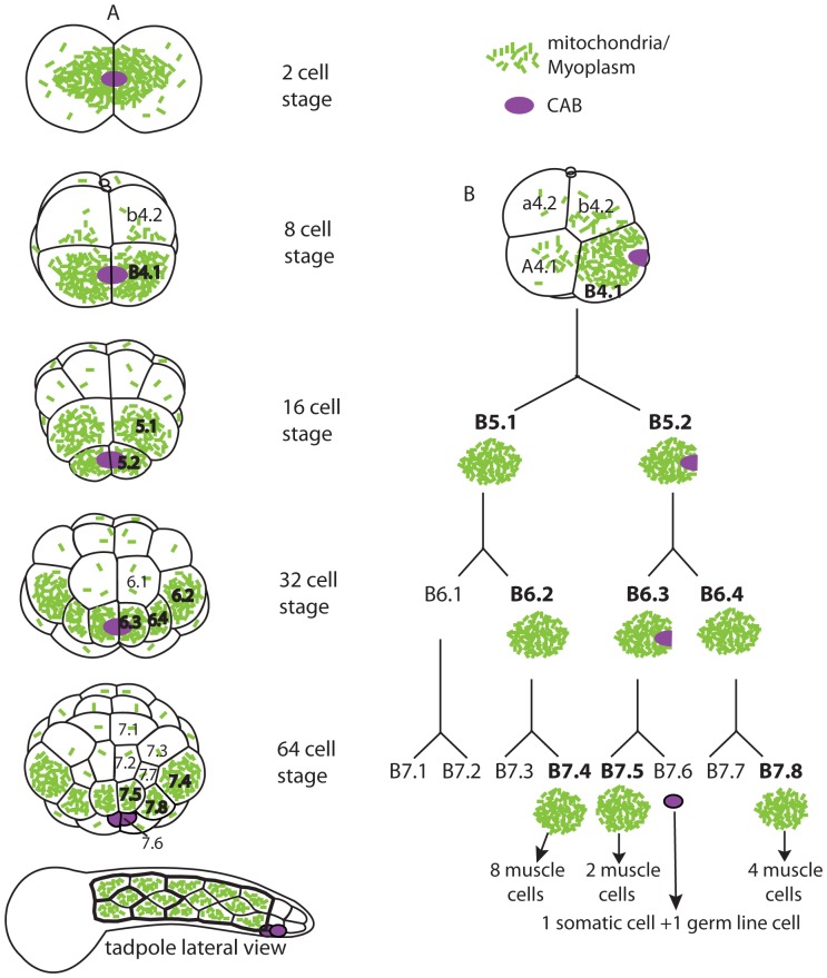
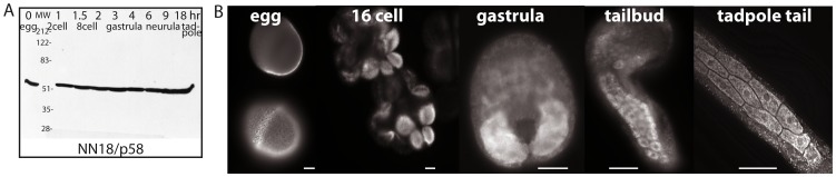
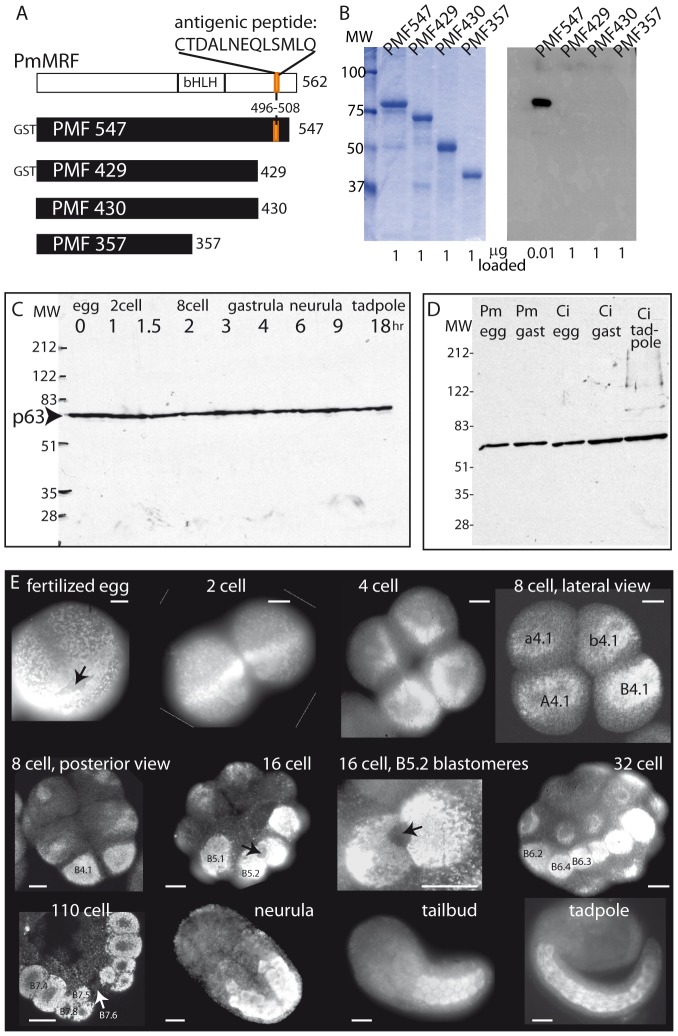
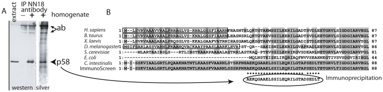
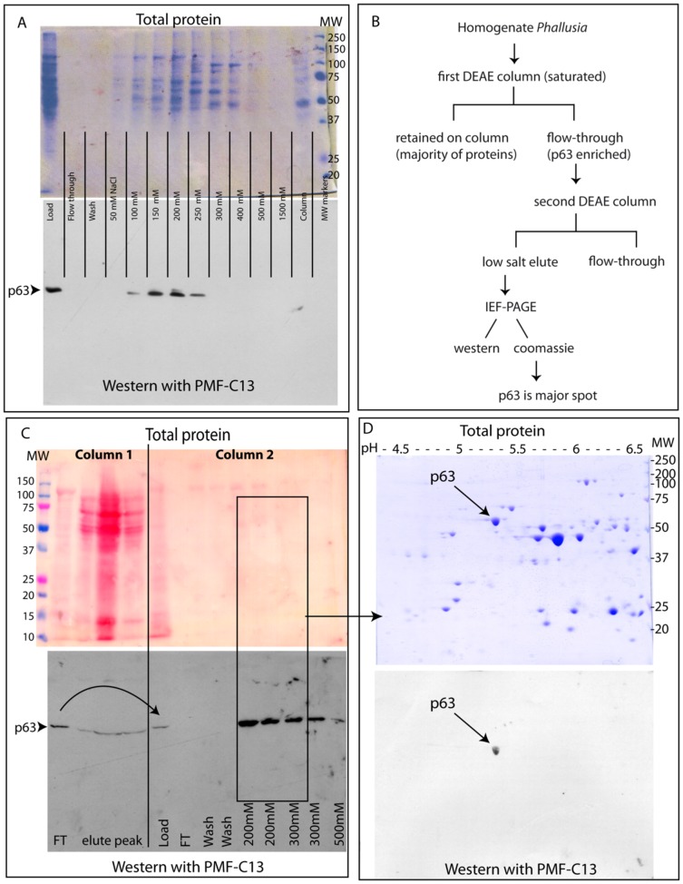
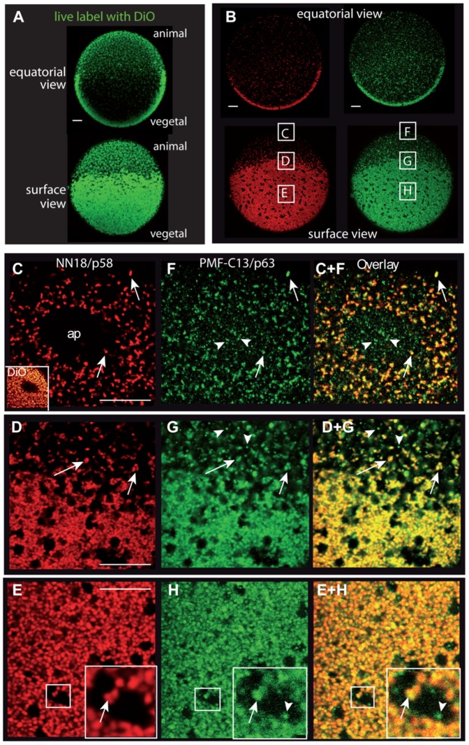
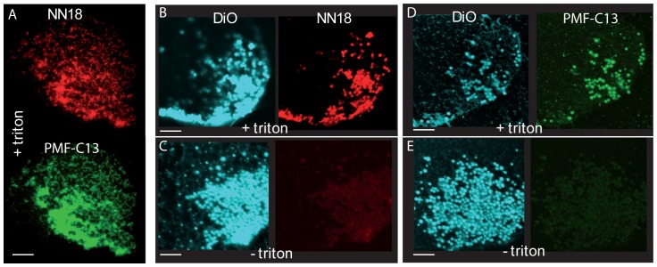
Use of antibodies is a cornerstone of biological studies and it is important to identify the recognized protein with certainty. Generally an antibody is considered specific if it labels a single band of the expected size in the tissue of interest, or has a strong affinity for the antigen produced in a heterologous system. The identity of the antibody target protein is rarely confirmed by purification and sequencing, however in many cases this may be necessary. In this study we sought to characterize the myoplasm, a mitochondria-rich domain present in eggs and segregated into tadpole muscle cells of ascidians (urochordates). The targeted proteins of two antibodies that label the myoplasm were purified using both classic immunoaffinity methods and a novel protein purification scheme based on sequential ion exchange chromatography followed by two-dimensional gel electrophoresis. Surprisingly, mass spectrometry sequencing revealed that in both cases the proteins recognized are unrelated to the original antigens. NN18, a monoclonal antibody which was raised against porcine spinal cord and recognizes the NF-M neurofilament subunit in vertebrates, in fact labels mitochondrial ATP synthase in the ascidian embryo. PMF-C13, an antibody we raised to and purified against PmMRF, which is the MyoD homolog of the ascidian Phallusia mammillata, in fact recognizes mitochondrial HSP60. High resolution immunolabeling on whole embryos and isolated cortices demonstrates localization to the inner mitochondrial membrane for both ATP synthase and HSP60. We discuss the general implications of our results for antibody specificity and the verification methods which can be used to determine unequivocally an antibody's target.
Conflict of interest statement
Figures
Similar articles
-
Identification of a cytoskeletal protein localized in the myoplasm of ascidian eggs: localization is modified during anural development.Development. 1991 Feb;111(2):425-36. doi: 10.1242/dev.111.2.425. Development. 1991. PMID: 1893869
-
Identification of two proteins associated with mammalian ATP synthase.Mol Cell Proteomics. 2007 Oct;6(10):1690-9. doi: 10.1074/mcp.M700097-MCP200. Epub 2007 Jun 17. Mol Cell Proteomics. 2007. PMID: 17575325
-
Localization and anchorage of maternal mRNAs to cortical structures of ascidian eggs and embryos using high resolution in situ hybridization.Methods Mol Biol. 2011;714:49-70. doi: 10.1007/978-1-61779-005-8_4. Methods Mol Biol. 2011. PMID: 21431734
-
Cell-cycle control in oocytes and during early embryonic cleavage cycles in ascidians.Int Rev Cell Mol Biol. 2012;297:235-64. doi: 10.1016/B978-0-12-394308-8.00006-6. Int Rev Cell Mol Biol. 2012. PMID: 22608561 Review.
-
Gene regulatory networks in the early ascidian embryo.Biochim Biophys Acta. 2009 Apr;1789(4):268-73. doi: 10.1016/j.bbagrm.2008.03.005. Epub 2008 Mar 30. Biochim Biophys Acta. 2009. PMID: 18424276 Review.
Cited by
-
Combined effect of cell geometry and polarity domains determines the orientation of unequal division.Elife. 2021 Dec 10;10:e75639. doi: 10.7554/eLife.75639. Elife. 2021. PMID: 34889186 Free PMC article.
-
Cellular remodeling and JAK inhibition promote zygotic gene expression in the Ciona germline.EMBO Rep. 2024 May;25(5):2188-2201. doi: 10.1038/s44319-024-00139-0. Epub 2024 Apr 22. EMBO Rep. 2024. PMID: 38649664 Free PMC article.
-
Actin Filament in the First Cell Cycle Contributes to the Determination of the Anteroposterior Axis in Ascidian Development.J Dev Biol. 2022 Feb 4;10(1):10. doi: 10.3390/jdb10010010. J Dev Biol. 2022. PMID: 35225963 Free PMC article.
-
Dynamic changes in the association between maternal mRNAs and endoplasmic reticulum during ascidian early embryogenesis.Dev Genes Evol. 2022 Feb;232(1):1-14. doi: 10.1007/s00427-021-00683-y. Epub 2021 Dec 18. Dev Genes Evol. 2022. PMID: 34921621 Free PMC article.
-
The Spindle Assembly Checkpoint Functions during Early Development in Non-Chordate Embryos.Cells. 2020 Apr 28;9(5):1087. doi: 10.3390/cells9051087. Cells. 2020. PMID: 32354040 Free PMC article.
References
-
- Deutsch EW, Ball CA, Berman JJ, Bova GS, Brazma A, et al. (2008) Minimum information specification for in situ hybridization and immunohistochemistry experiments (MISFISHIE). Nat Biotechnol 26: 305–312 doi:10.1038/nbt1391. - DOI - PMC - PubMed
-
- True LD (2008) Quality control in molecular immunohistochemistry. Histochem Cell Biol 130: 473–480 doi:10.1007/s00418-008-0481-0. - DOI - PMC - PubMed
-
- Saper CB (2009) A guide to the perplexed on the specificity of antibodies. J Histochem Cytochem 57: 1–5 doi:10.1369/jhc.2008.952770. - DOI - PMC - PubMed
-
- Bordeaux J, Welsh A, Agarwal S, Killiam E, Baquero M, et al. (2010) Antibody validation. BioTechniques 48: 197–209 doi:10.2144/000113382. - DOI - PMC - PubMed
-
- Burry RW (2011) Controls for immunocytochemistry: an update. J Histochem Cytochem 59: 6–12 doi:10.1369/jhc.2010.956920. - DOI - PMC - PubMed
Publication types
MeSH terms
Substances
LinkOut - more resources
Full Text Sources
Other Literature Sources
Research Materials
Miscellaneous
