. 2013;8(1):e53301.
doi: 10.1371/journal.pone.0053301. Epub 2013 Jan 9.

Investigate pathogenic mechanism of TXNDC5 in rheumatoid arthritis

Affiliations

Investigate pathogenic mechanism of TXNDC5 in rheumatoid arthritis

Lin Wang et al. PLoS One. 2013.

Abstract

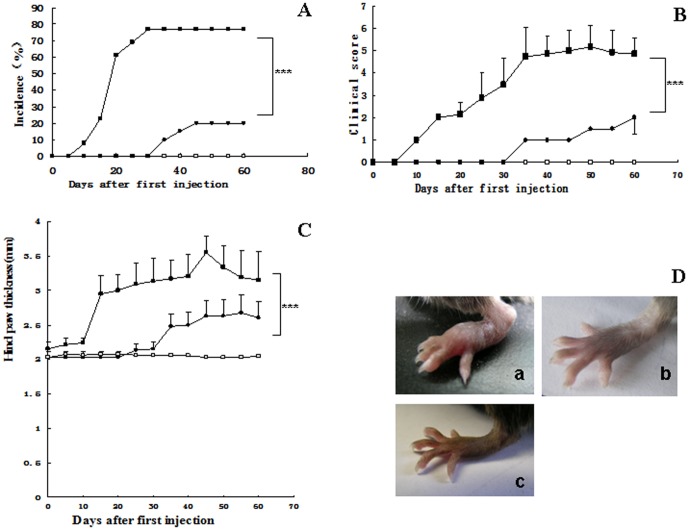
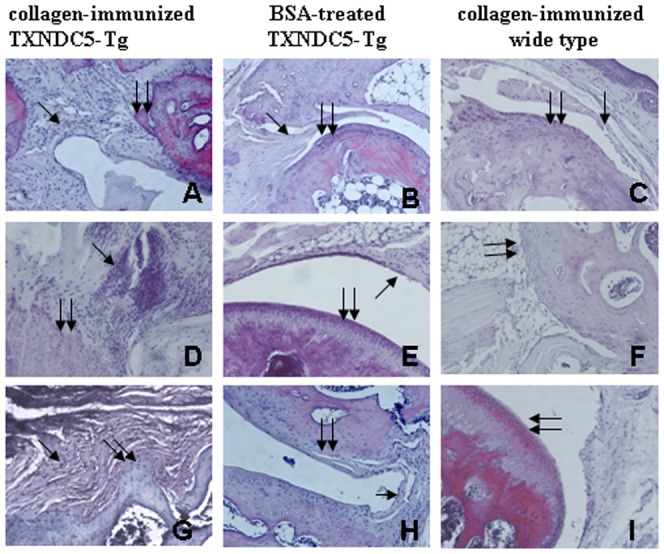
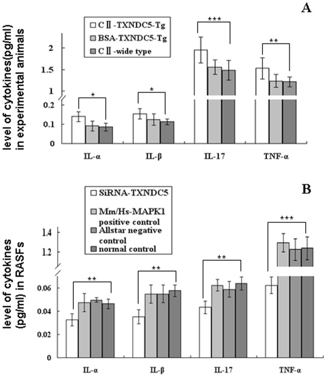
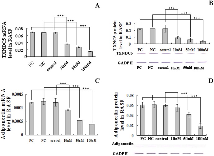
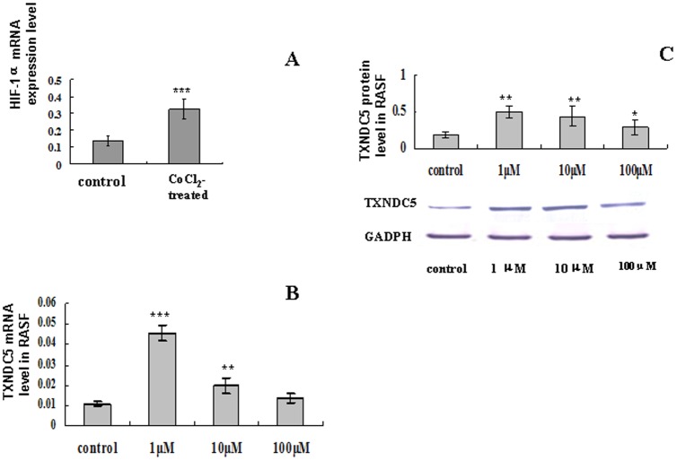
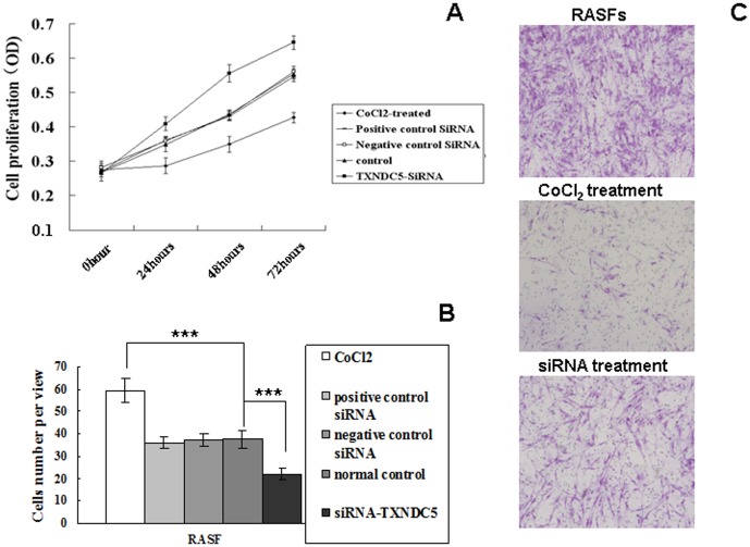
Hypoxia stimulates synovial hypoperfusion in rheumatoid arthritis (RA). TXNDC5 stimulates cellular proliferation in hypoxic conditions. We previously detected increased TXNDC5 expression in synovial tissues and blood from RA patients and demonstrated that the gene encoding TXNDC5 increased RA risk. The present study investigated the pathogenic roles of TXNDC5 in RA. Transgenic mice that over-expressed TXNDC5 (TXNDC5-Tg) were generated using C57BL/6J mice and treated with bovine collagen II to induce arthritis (CIA). Synovial fibroblasts from RA patients (RASFs) were cultured and incubated with TXNDC5-siRNA or CoCl(2), a chemical that induces hypoxia. CIA was observed in 80% of the TXNDC5-Tg, but only 20% of the wild-type mice (WT) developed CIA. The clinical arthritis scores reached 5 in the TXNDC5-Tg, but this index only reached 2 in the control mice. CIA TXNDC5-Tg exhibited clear pannus proliferation and bone erosion in joint tissues. A significant increase in CD4 T cells was observed in the thymus and spleen of TXNDC5-Tg during CIA. Serum levels of anti-collagen II IgG, IgG1 and IgG2a antibodies were significantly elevated in the mice. Increased cell proliferation, cell migration and TXNDC5 expression were observed in RASFs following incubation with 1 µM CoCl(2). However, this effect was diminished when TXNDC5 expression was inhibited with 100 nM siRNA. TNF-alpha, IL-1α, IL-1β and IL-17 levels were significantly increased in the blood of TXNDC5-Tg mice, but the levels of these cytokines declined in the supernatant of RASFs that were treated with TXNDC5 siRNA. The expression of adiponectin, a cytokine-like mediator, decreased significantly in RASFs following TXNDC5 siRNA treatment. These results suggest that TXNDC5-over-expressing mice were susceptible to CIA. This study also suggests that hypoxia induced TXCNDC5 expression, which contributed to adiponectin expression, cytokine production and the cellular proliferation and migration of fibroblasts in RA.


Conflict of interest statement

Competing Interests: The authors have declared that no competing interests exist.

Figures

Figure 1
Figure 1. Clinical arthritis symptoms in collagen-injected TXNDC5-Tg mice.
The incidence of arthritis (A), the hind paw clinical score (B) and the hind paw thickness (C) in TXNDC5-Tg (filled squares, n = 15) and WT (filled circles, n = 10) mice were measured at various time points following the first injection. A separate group of TXNDC5-Tg mice received BSA, and these mice were used as normal controls (empty squares, n = 10). (D) The symptoms of CIA were observed in the hind paws of these collagen-treated mice. a) represents collagen-injected TXNDC5-Tg, b) represents BSA-treated TXNDC5-Tg, c) represents collagen-injected wild type. * = p<0.05, ** = p<0.01, *** = p<0.001.
Figure 2
Figure 2. Histochemical examination of collagen-induced arthritis in experimental mice.
The tissue structures of the paw (A–C), knee (E–F) and ankle (G–I) were histochemically examined. Synovial hyperplasia and inflammation, cartilage destruction and bone resorption with pannus formation were observed in the arthritic joints of the collagen-treated TXNDC5-Tg mice (n = 5) but not in WT (n = 5) and TXNDC5-Tg mice treated with BSA (n = 5). Single arrow represents synovial membrane, and double arrow indicates joint cartilage. Magnification: 200×.
Figure 3
Figure 3. Measurement of cytokine levels using ELISA.
(A) IL-1α, IL-1β, TNF-α and IL-17 levels in the sera of TXNDC5-Tg (n = 5) and WT mice following the injection of bovine collagen II (n = 5) and TXNDC5-Tg mice injected with BSA (n = 5). TXNDC5-Tg mice exhibited higher sera levels of TNF-α, IL-1α, IL-1β and IL-17 than wild type mice with treatment. (B) IL-1α, IL-1β, TNF-α and IL-17 levels in the supernatant of TXNDC5 siRNA (100 nM)-treated RASFs (n = 15). Parallel experiments with Mm/Hs-MAPK1 siRNA and Allstar siRNA were used as controls. RASFs without siRNA were also used as controls. TNF-α, IL-1α, IL-1β and IL-17 levels decreased significantly in RASF supernatants following treatment with TXNDC5 siRNA compared to the normal controls without the siRNA treatment. * = p<0.05, ** p<0.01, *** = p<0.001.
Figure 4
Figure 4. TXNDC5 and adiponectin expression in TXNDC5 siRNA-treated RASFs.
The RASFs were transiently transfected with TXNDC5 siRNA (n = 15), and the effects of TXNDC5 knockdown were determined at the mRNA (A) and protein levels (B) using real-time PCR and Western blotting, respectively. The effect of inhibiting TXNDC5 expression on adiponectin expression was also determined using real-time PCR (C) and Western blotting (D). The mRNA levels of TXNDC5 and adiponectin were normalized to β-actin. The protein levels of TXNDC5 and adiponectin were normalized to GAPDH. TXNDC5 and adiponectin levels were not significantly changed between the positive control, the negative control and the cells without siRNA treatment. PC indicates the positive control, and NC indicates the negative control. * = p<0.05, ** p<0.01, *** = p<0.001.
Figure 5
Figure 5. TXNDC5 expression in RASFs (n = 15) following treatment with different concentrations of CoCl2.
(A) HIF-1α expression was detected in RASFs prior to and following 1 µM CoCl2 treatment. Real-time PCR and Western blotting analyses were used to measure TXNDC5 mRNA (B) and protein expression (C). * = p<0.05, ** p<0.01, *** = p<0.001.
Figure 6
Figure 6. Cell proliferation and cell invasion in RASFs (n = 15) treated with CoCl2 and TXNDC5-siRNA.
(A) The cell viability of RASFs was determined using an MTT assay. (B) The invasive ability of RASFs was examined using a 2-compartment transwell system. The average number of cells that invaded through the filter was quantified. (C) Crystal violet staining of the lower surface filters revealed the cells that passed through the filter and attached to the lower side of the filter (100×). The data were obtained from three independent experiments. * = p<0.05, ** p<0.01, *** = p<0.001.

Similar articles

Cited by

References

    1. Nakamura H (2004) Thioredoxin as a key molecule in redox signaling. Antioxid Redox Signa l6: 15–17. - PubMed
    1. Sullivan DC, Huminiecki L, Moore JW, Boyle JJ, Poulsom R, et al. (2003) EndoPDI, a novel protein-disulfide isomerase-like protein that is preferentially expressed in endothelial cells acts as a stress survival factor. J Biol Chem 278: 47079–47088. - PubMed
    1. Muz B, Khan MN, Kiriakidis S, Paleolog EM (2009) The role of hypoxia and HIF-α dependent signalling events in rheumatoid arthritis. Arthritis Res Ther 11: R201. - PMC - PubMed
    1. Chang X, Cui Y, Zong M, Zhao Y, Yan X, et al. (2009) Identification of proteins with increased expression in rheumatoid arthritis synovial tissues. J Rheumatol 36: 872–880. - PubMed
    1. Chang X, Zhao Y, Yan X, Pan J, Fang K, et al. (2011) Investigating a pathogenic role for TXNDC5 in rheumatoid arthritis. Arthritis Res Ther 13: R124. - PMC - PubMed

Publication types

MeSH terms