

Mapping the complex morphology of cell interactions with nanowire substrates using FIB-SEM
- PMID: 23326412
- PMCID: PMC3541134
- DOI: 10.1371/journal.pone.0053307
Mapping the complex morphology of cell interactions with nanowire substrates using FIB-SEM
Abstract
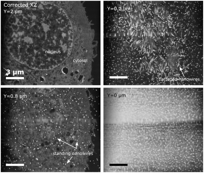
Using high resolution focused ion beam scanning electron microscopy (FIB-SEM) we study the details of cell-nanostructure interactions using serial block face imaging. 3T3 Fibroblast cellular monolayers are cultured on flat glass as a control surface and on two types of nanostructured scaffold substrates made from silicon black (Nanograss) with low- and high nanowire density. After culturing for 72 hours the cells were fixed, heavy metal stained, embedded in resin, and processed with FIB-SEM block face imaging without removing the substrate. The sample preparation procedure, image acquisition and image post-processing were specifically optimised for cellular monolayers cultured on nanostructured substrates. Cells display a wide range of interactions with the nanostructures depending on the surface morphology, but also greatly varying from one cell to another on the same substrate, illustrating a wide phenotypic variability. Depending on the substrate and cell, we observe that cells could for instance: break the nanowires and engulf them, flatten the nanowires or simply reside on top of them. Given the complexity of interactions, we have categorised our observations and created an overview map. The results demonstrate that detailed nanoscale resolution images are required to begin understanding the wide variety of individual cells' interactions with a structured substrate. The map will provide a framework for light microscopy studies of such interactions indicating what modes of interactions must be considered.
Conflict of interest statement
Figures


Similar articles
-
Combining serial block face and focused ion beam scanning electron microscopy for 3D studies of rare events.Methods Cell Biol. 2019;152:87-101. doi: 10.1016/bs.mcb.2019.03.014. Epub 2019 May 7. Methods Cell Biol. 2019. PMID: 31326028
-
3D imaging of cells and tissues by focused ion beam/scanning electron microscopy (FIB/SEM).Methods Mol Biol. 2013;950:275-92. doi: 10.1007/978-1-62703-137-0_16. Methods Mol Biol. 2013. PMID: 23086881
-
FIB-SEM imaging properties of Drosophila melanogaster tissues embedded in Lowicryl HM20.J Microsc. 2019 Feb;273(2):91-104. doi: 10.1111/jmi.12764. Epub 2018 Nov 12. J Microsc. 2019. PMID: 30417390
-
Volume microscopy in biology: FIB-SEM tomography.Tissue Cell. 2019 Apr;57:123-128. doi: 10.1016/j.tice.2018.09.006. Epub 2018 Sep 26. Tissue Cell. 2019. PMID: 30385054 Review.
-
Focused ion beam scanning electron microscopy in biology.J Microsc. 2014 Jun;254(3):109-14. doi: 10.1111/jmi.12127. Epub 2014 Apr 7. J Microsc. 2014. PMID: 24707797 Review.
Cited by
-
Interfacing Cells with Vertical Nanoscale Devices: Applications and Characterization.Annu Rev Anal Chem (Palo Alto Calif). 2018 Jun 12;11(1):101-126. doi: 10.1146/annurev-anchem-061417-125705. Epub 2018 Mar 23. Annu Rev Anal Chem (Palo Alto Calif). 2018. PMID: 29570360 Free PMC article. Review.
-
Cellular uptake and dynamics of unlabeled freestanding silicon nanowires.Sci Adv. 2016 Dec 16;2(12):e1601039. doi: 10.1126/sciadv.1601039. eCollection 2016 Dec. Sci Adv. 2016. PMID: 28028534 Free PMC article.
-
Adhesion Stabilized en Masse Intracellular Electrical Recordings from Multicellular Assemblies.Nano Lett. 2019 May 8;19(5):3244-3255. doi: 10.1021/acs.nanolett.9b00784. Epub 2019 Apr 12. Nano Lett. 2019. PMID: 30950627 Free PMC article.
-
FIB-SEM imaging of carbon nanotubes in mouse lung tissue.Anal Bioanal Chem. 2014 Jun;406(16):3863-73. doi: 10.1007/s00216-013-7566-x. Epub 2014 Jan 22. Anal Bioanal Chem. 2014. PMID: 24448971 Free PMC article.
-
High Aspect Ratio and Light-Sensitive Micropillars Based on a Semiconducting Polymer Optically Regulate Neuronal Growth.ACS Appl Mater Interfaces. 2021 May 26;13(20):23438-23451. doi: 10.1021/acsami.1c03537. Epub 2021 May 13. ACS Appl Mater Interfaces. 2021. PMID: 33983012 Free PMC article.
References
-
- McKnight TE, Melechko AV, Griffin GD, Guillorn MA, Merkulov VI, et al. (2003) Intracellular integration of synthetic nanostructures with viable cells for controlled biochemical manipulation. Nanotechnology 14: 551–556 doi:10.1088/0957–4484/14/5/313 - DOI
-
- Shalek AK, Robinson JT, Karp ES, Lee JS, Ahn D-R, et al. (2010) Vertical silicon nanowires as a universal platform for delivering biomolecules into living cells. Proceedings of the National Academy of Sciences 107: 1870–1875 doi:10.1073/pnas.0909350107 - DOI - PMC - PubMed
-
- Bittermann AG, Burkhardt C, Hall H (2009) Imaging of Cell-to-Material Interfaces by SEM after in situ Focused Ion Beam Milling on Flat Surfaces and Complex 3D-Fibrous Structures. Adv Eng Mater 11: B182–B188 doi:10.1002/adem.200900080 - DOI
-
- Łopacińska JM, Grădinaru C, Wierzbicki R, Købler C, Schmidt MS, et al. (2012) Cell motility, morphology, viability and proliferation in response to nanotopography on silicon black. Nanoscale 4: 3739 doi:10.1039/c2nr11455k - DOI - PubMed
Publication types
MeSH terms
Substances
LinkOut - more resources
Full Text Sources
Other Literature Sources
