. 2013;8(1):e53326.
doi: 10.1371/journal.pone.0053326. Epub 2013 Jan 11.

Structure-activity relationships of the antimicrobial peptide arasin 1 - and mode of action studies of the N-terminal, proline-rich region

Affiliations

Structure-activity relationships of the antimicrobial peptide arasin 1 - and mode of action studies of the N-terminal, proline-rich region

Victoria S Paulsen et al. PLoS One. 2013.

Abstract

Arasin 1 is a 37 amino acid long proline-rich antimicrobial peptide isolated from the spider crab, Hyas araneus. In this work the active region of arasin 1 was identified through structure-activity studies using different peptide fragments derived from the arasin 1 sequence. The pharmacophore was found to be located in the proline/arginine-rich NH(2) terminus of the peptide and the fragment arasin 1(1-23) was almost equally active to the full length peptide. Arasin 1 and its active fragment arasin 1(1-23) were shown to be non-toxic to human red blood cells and arasin 1(1-23) was able to bind chitin, a component of fungal cell walls and the crustacean shell. The mode of action of the fully active N-terminal arasin 1(1-23) was explored through killing kinetic and membrane permeabilization studies. At the minimal inhibitory concentration (MIC), arasin 1(1-23) was not bactericidal and had no membrane disruptive effect. In contrast, at concentrations of 5×MIC and above it was bactericidal and interfered with membrane integrity. We conclude that arasin 1(1-23) has a different mode of action than lytic peptides, like cecropin P1. Thus, we suggest a dual mode of action for arasin 1(1-23) involving membrane disruption at peptide concentrations above MIC, and an alternative mechanism of action, possibly involving intracellular targets, at MIC.


Conflict of interest statement

Competing Interests: The authors have declared that no competing interests exist.

Figures

Figure 1
Figure 1. Overview of synthetic arasin 1 and its deletion peptides.
Shaded letters indicate characteristic residues for arasin (Cys: black) and for proline-rich AMPs (Pro, Arg: grey). Acm = Acetamidomethyl. Predicted disulphide bridges are indicated for arasin 1 .
Figure 2
Figure 2. CD spectra of arasin 1(1–23) (A) and arasin 1 (B).
Peptides were analyzed at a final concentration of 20 µM in 10 mM sodium phosphate buffer pH 7.0 (solid line) or with the addition of 10 mM SDS (dotted lines). Spectra are the smoothed means of at least two measurements, each composed of three different scans.
Figure 3
Figure 3. Killing kinetics of E. coli cells treated with arasin 1(1–23) or cecropin P1.
Colony forming units (CFU) of E. coli HB101 subjected to peptide treatment or water (negative control) are shown. Sample aliquots were withdrawn and washed in high salt solution at the time points indicated and subsequently plated for colony counts. Data are expressed as the average number of colony forming cells ± S.D. for three independent experiments.
Figure 4
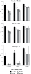
Figure 4. Effect of arasin 1(1–23), PR-39(1–26) and cecropin P1 on E. coli viability.
Relative light units (rlu) produced by E.coli HB101, constitutively expressing the luxCDABE operon are shown, after AMP treatment at different concentrations. Light emission is shown relative to the untreated control for the 4 selected incubation times. The mean of three independent measurements is indicated ± S.D. MIC against E. coli HB101 is 4, 1 and 1 µM for arasin 1(1–23), PR-39(1–26) and cecropin P1, respectively.
Figure 5
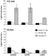
Figure 5. Effect of arasin 1(1–23), PR-39(1–26) and cecropin P1 on E. coli membrane integrity after 0.5 and 10 min incubation.
Light induction of luciferase expressing E. coli HB101 after AMP treatment at different concentrations is shown. The data presented are relative to the untreated control for the two selected incubation times. The mean of three independent measurements is indicated ± S.D. Note that for the highly membrane active peptide cecropin P1 light emission for the highest concentrations already peaks before the first measurement. MIC against E. coli HB101 is 4, 1 and 1 µM for arasin 1(1–23), PR-39(1–26) and cecropin P1, respectively.
Figure 6
Figure 6. Bacterial membrane integrity after treatment with arasin 1(1–23) or cecropin P1.
Percentage of fluorescent cells (PI-positive) measured by flow cytometry after incubation of E. coli HB101 cells with 4, 20, and 40 µM arasin 1(1–23) or 4 µM cecropin P1 is shown. The background level of permeabilized cells, obtained using untreated samples, was always lower than 3% and was subtracted to the corresponding peptide-treated sample. Data are expressed as the average of % PI-positive cells ± S.D. for four independent experiments.
Figure 7
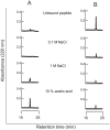
Figure 7. Chitin binding capacity of (A) arasin 1(1–23) and (B) arasin 1(20–37)-Acm.
Fifty micrograms of peptides were incubated with 40 mg chitin, and subsequently washed with 0.1 M, 1 M NaCl, and hot (95°C) 10% acetic acid. The obtained supernatants were subjected to RP-HPLC on a C18 column. The resulting chromatograms show unbound material and peptides released from chitin by the different washing solutions, respectively.

Similar articles

Cited by

References

    1. Hancock RE, Diamond G (2000) The role of cationic antimicrobial peptides in innate host defences. Trends Microbiol 8: 402–410. - PubMed
    1. Peters BM, Shirtliff ME, Jabra-Rizk MA (2010) Antimicrobial peptides: primeval molecules or future drugs? PLoS Pathog 6: e1001067. - PMC - PubMed
    1. Wang G, Li X, Wang Z (2009) APD2: the updated antimicrobial peptide database and its application in peptide design. Nucleic Acids Res 37: D933–937. - PMC - PubMed
    1. Shai Y (1999) Mechanism of the binding, insertion and destabilization of phospholipid bilayer membranes by alpha-helical antimicrobial and cell non-selective membrane-lytic peptides. Biochim Biophys Acta 1462: 55–70. - PubMed
    1. Shai Y (2002) Mode of action of membrane active antimicrobial peptides. Biopolymers 66: 236–248. - PubMed

Publication types

MeSH terms

LinkOut - more resources