. 2013;8(1):e53414.
doi: 10.1371/journal.pone.0053414. Epub 2013 Jan 9.

Intestinal T-cell responses in celiac disease - impact of celiac disease associated bacteria

Affiliations

Intestinal T-cell responses in celiac disease - impact of celiac disease associated bacteria

Veronika Sjöberg et al. PLoS One. 2013.

Abstract

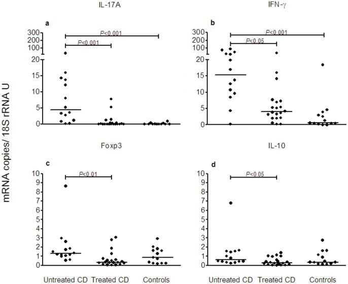
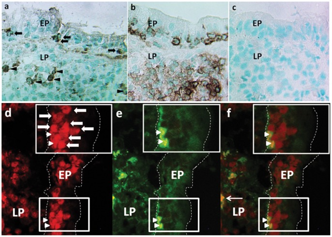
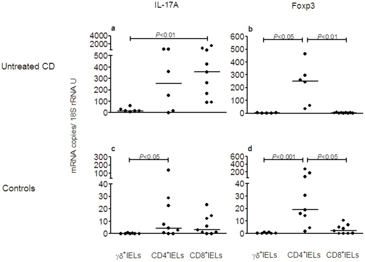
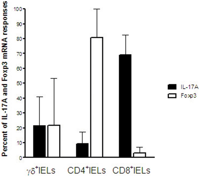
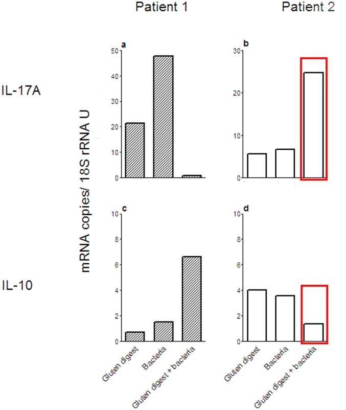
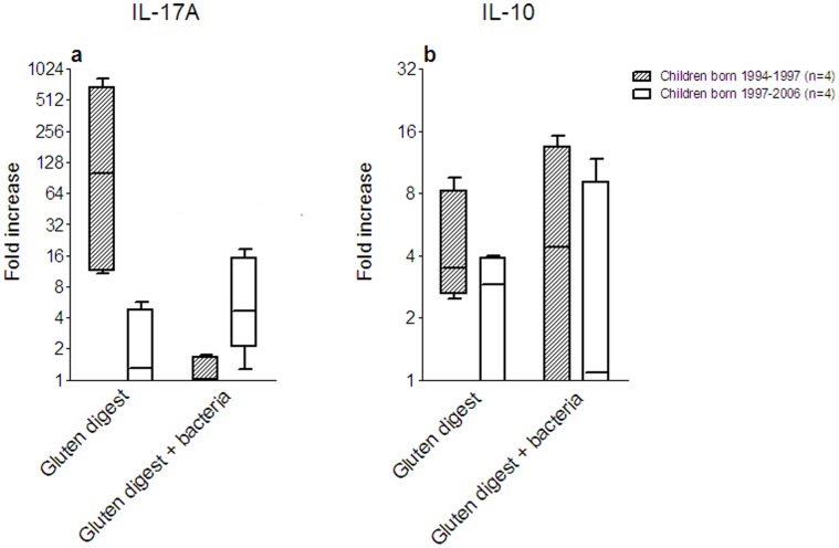
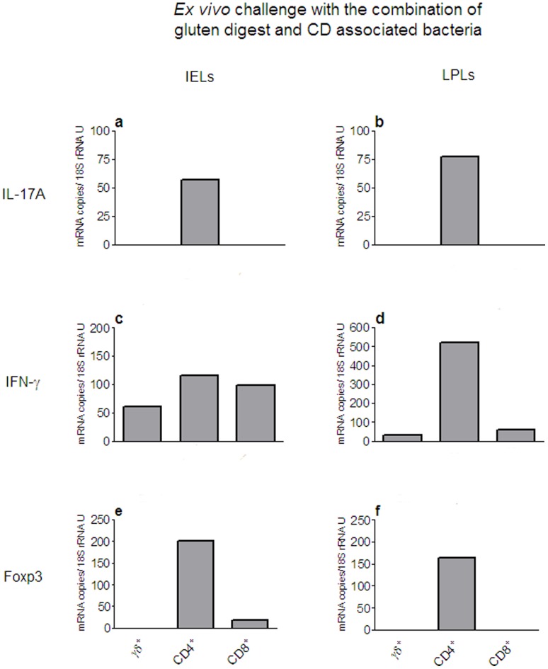
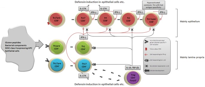
A hallmark of active celiac disease (CD), an inflammatory small-bowel enteropathy caused by permanent intolerance to gluten, is cytokine production by intestinal T lymphocytes. Prerequisites for contracting CD are that the individual carries the MHC class II alleles HLA-DQ2 and/or HLA-DQ8 and is exposed to gluten in the diet. Dysbiosis in the resident microbiota has been suggested to be another risk factor for CD. In fact, rod shaped bacteria adhering to the small intestinal mucosa were frequently seen in patients with CD during the "Swedish CD epidemic" and bacterial candidates could later be isolated from patients born during the epidemic suggesting long-lasting changes in the gut microbiota. Interleukin-17A (IL-17A) plays a role in both inflammation and anti-bacterial responses. In active CD IL-17A was produced by both CD8(+) T cells (Tc17) and CD4(+) T cells (Th17), with intraepithelial Tc17 cells being the dominant producers. Gluten peptides as well as CD associated bacteria induced IL-17A responses in ex vivo challenged biopsies from patients with inactive CD. The IL-17A response was suppressed in patients born during the epidemic when a mixture of CD associated bacteria was added to gluten, while the reverse was the case in patients born after the epidemic. Under these conditions Th17 cells were the dominant producers. Thus Tc17 and Th17 responses to gluten and bacteria seem to pave the way for the chronic disease with interferon-γ-production by intraepithelial Tc1 cells and lamina propria Th1 cells. The CD associated bacteria and the dysbiosis they might cause in the resident microbiota may be a risk factor for CD either by directly influencing the immune responses in the mucosa or by enhancing inflammatory responses to gluten.


Conflict of interest statement

Competing Interests: The authors have declared that no competing interests exist.

Figures

Figure 1
Figure 1. IL-17A mRNA levels are elevated in patients with active CD.
Expression levels of mRNAs for IL-17A (a), IFN-γ (b), Foxp3 (c) and IL-10 (d) were determined in freshly taken biopsies from the proximal small intestine of pediatric CD patients with untreated, active disease (Untreated CD), CD patients on gluten-free diet with inactive disease (Treated CD) and clinical controls (Controls) using qRT-PCR. Amounts of cytokine and Foxp3 mRNAs were normalized to the 18S rRNA content in each sample. Dots depict expression levels in individual samples. Horizontal bars indicate median values. P-values of statistically significant differences are depicted.
Figure 2
Figure 2. Tc17 cells are present in the small intestinal mucosa of patients with active CD.
Immunoperoxidase (ac) and immunofluorescence (df) staining of the small intestinal mucosa of patients with untreated CD. (ac) sequential sections of intestinal mucosa of one CD patient with active disease stained with anti-IL-17A mAb (a) showing cells with cytoplasmic staining for IL-17A both within the epithelium (black arrows) and in the LP (black arrowheads), suggesting IL-17A secretion at both locations; (b), positive control, i.e. section stained with anti-CD45 mAbs identifying all leukocytes; (c), negative control, i.e. section incubated with concentration and isotype matched irrelevant mAb. (df) a cryosection of intestinal mucosa of one CD patient with active disease stained in two-color immunofluorescence. (d), CD8+ cells stained in red. (e), IL-17A+ cells stained in green. (f), overlay of (d) and (e) showing yellow IL-17A+CD8+ cells. The box in (d) encloses a cluster of 9 CD8+ IELs, two of them are also positive for IL-17A (e, f). Thick white arrows depict CD8 single positive IELs. White arrowheads depict CD8/IL-17A double positive IELs. Thin white arrow depicts a CD8/IL-17A double positive LPL. Original magnification, X 220 (ac) and X 400 (df). EP indicates epithelium and LP lamina propria. Dotted white lines in d–f indicate the basal lamina and the apical surface of the epithelium. Smaller boxes in (df) indicate the area enlarged in the upper right corners.
Figure 3
Figure 3. High levels of IL-17A mRNA in intraepithelial Tc17 and Th17 cells in active CD.
Expression levels of IL-17A (a, c) and Foxp3 (b, d) mRNAs were determined in TCR-γδ+ IELs (γδ+IELs), CD4+ TCR-γδ IELs (CD4+IELs), and CD8+ CD4 TCR-γδ IELs (CD8+IELs) retrieved from IELs isolated from intestinal biopsies of patients with untreated CD (Untreated CD) and controls (Controls). Dots depict expression levels for the indicated mRNA species in individual samples. Horizontal bars indicate median values. P-values of statistically significant differences are depicted.
Figure 4
Figure 4. Tc17 cells contribute most of the IL-17A mRNA in the intestinal epithelium in active CD.
TCR-γδ+IELs (γδ+IELs), CD4+IELs and CD8+IELs were sequentially retrieved from small intestinal biopsies of 4 patients with untreated CD and analyzed for IL-17A mRNA and Foxp3 mRNA. Results are expressed as relative contribution in percent to the total amount of mRNA of the indicated species in the γδ+IEL, CD4+IEL and CD8+IEL subsets of each individual. Bars indicate mean and whiskers 1 SD. Filled bars, IL-17A mRNA; Open bars, Foxp3 mRNA.
Figure 5
Figure 5. The IL-17A responses of ex vivo challenged biopsies show two different patterns.
Expression levels of IL-17A (ab) and IL-10 (cd) mRNAs were determined in biopsies from treated CD patients after challenge ex vivo with gluten digest (Gluten digest), a mixture of CD associated bacteria (Bacteria), a combination of gluten digest and the mixture of CD associated bacteria (Gluten digest+bacteria), or incubated with medium alone. Bars indicate the mRNA expression level for the indicated stimulant of each biopsy after subtraction of the mRNA level in the medium control for Patient #1, born 1996 (a, c), and Patient #2, born 2006 (b, d). Determinations were done in triplicates. Red boxes in (b) and (d) indicate the responses to challenge with the combination of gluten digest and a mixture of CD associated bacteria in the whole biopsy before fractionation into T cell subsets for which the results are shown in Figure 7.
Figure 6
Figure 6. CD associated bacteria suppress the IL-17A response to gluten in children born during the “epidemic”.
IL-17A (a) and IL-10 (b) responses to ex vivo challenge with gluten digest alone (Gluten digest) and the combination of gluten digest and a mixture of CD associated bacteria (Gluten digest+bacteria) shown as fold increase calculated as the ratio over the individual medium control for patients #1 through 8 and grouped according to the IL-17A response to challenge with gluten digest alone. Horizontal bars indicate medians, boxes indicate the 25th to 75th IQR, and whiskers indicate the range. Filled boxes and bars: Treated CD patients with strong IL-17A responses to challenge with gluten digest alone, all born during the Swedish CD epidemic. Open boxes and bars: Treated CD patents with weak IL-17A responses to challenge with gluten digest alone, all born after the epidemic. n = 4 in each group.
Figure 7
Figure 7. Ex vivo challenge with gluten digest combined with CD associated bacteria provokes a Th17 response.
Expression levels of IL-17A (a, b), IFN-γ (c, d), and Foxp3 (e, f) mRNAs determined in TCR-γδ+ (γδ+)-, CD4+ TCR-γδ (CD4+)-, and CD8+ CD4 TCR-γδ (CD8+)-cells retrieved from IELs and LPLs isolated from an intestinal biopsy of patient #2 in Figure 5 after challenge ex vivo with a combination of gluten digest and a mixture of CD associated bacteria. The IL-17A and IL-10 responses in the whole biopsy are indicated by red boxes in Figure 5 (b) and (d). Bars indicate the mRNA expression level of each T cell subtype. Determinations were done in triplicates.
Figure 8
Figure 8. Hypothetical scenario of the immune situation in the small intestinal mucosa during the inflammatory reaction.
The reaction to challenge at the mucosal lining with gluten peptides and components of CD associated bacteria induces at process during which the intraepithelial Tc17 cells (red) become activated and gradually transform to hyperactivated cytotoxic T lymphocytes with lost antigen specificity. IFN-γ drives the development both in an autocrine manner with Tc17 cells converting to Tc1 cells and paracrine manner through Th1 cells (green) specifically stimulated by gluten peptides presented on HLA-DQ2/DQ8. Parallel activation of Tregs (violet) occurs. These Tregs secrete IL-10 that down-regulates Th17 (blue) and Th1 cells but the Tc17 cells are resistant to down-regulation. IL-17A secreted by Tc17 and Th17 cells upon activation by gluten peptides and/or bacterial components promotes both inflammation and an antibacterial response including for example defensin production by epithelial cells.

Similar articles

Cited by

References

    1. Sollid LM (2002) Coeliac disease: dissecting a complex inflammatory disorder. Nat Rev Immunol 2: 647–655. - PubMed
    1. Tack GJ, Verbeek WH, Schreurs MW, Mulder CJ (2010) The spectrum of celiac disease: epidemiology, clinical aspects and treatment. Nat Rev Gastroenterol Hepatol 7: 204–213. - PubMed
    1. Di Sabatino A, Corazza GR (2009) Coeliac disease. Lancet 373: 1480–1493. - PubMed
    1. Forsberg G, Hernell O, Melgar S, Israelsson A, Hammarström S, et al. (2002) Paradoxical coexpression of proinflammatory and down-regulatory cytokines in intestinal T cells in childhood celiac disease. Gastroenterology 123: 667–678. - PubMed
    1. Sollid LM, Khosla C (2011) Novel therapies for coeliac disease. J Intern Med 269: 604–613. - PMC - PubMed

Publication types

MeSH terms