

3,3'-Diindolylmethane protects against cardiac hypertrophy via 5'-adenosine monophosphate-activated protein kinase-α2
- PMID: 23326427
- PMCID: PMC3541184
- DOI: 10.1371/journal.pone.0053427
3,3'-Diindolylmethane protects against cardiac hypertrophy via 5'-adenosine monophosphate-activated protein kinase-α2
Abstract
Purpose: 3,3'-Diindolylmethane (DIM) is a natural component of cruciferous plants. It has strong antioxidant and anti-angiogenic effects and promotes the apoptosis of a variety of tumor cells. However, little is known about the critical role of DIM on cardiac hypertrophy. In the present study, we investigated the effects of DIM on cardiac hypertrophy.
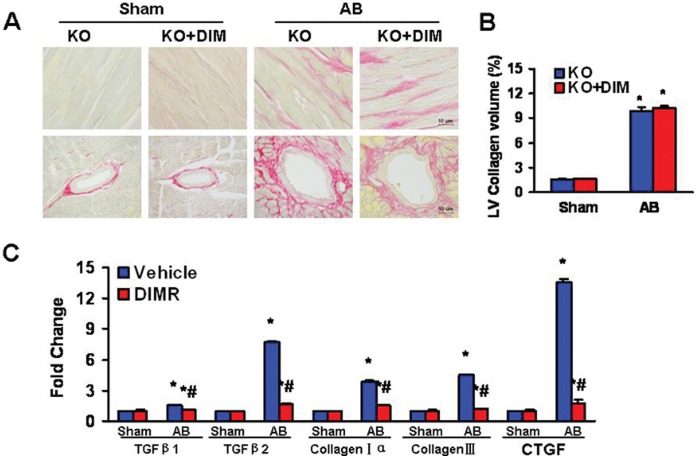
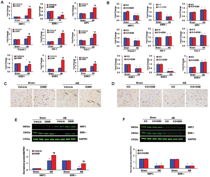
Methods: Multiple molecular techniques such as Western blot analysis, real-time PCR to determine RNA expression levels of hypertrophic, fibrotic and oxidative stress markers, and histological analysis including H&E for histopathology, PSR for collagen deposition, WGA for myocyte cross-sectional area, and immunohistochemical staining for protein expression were used.
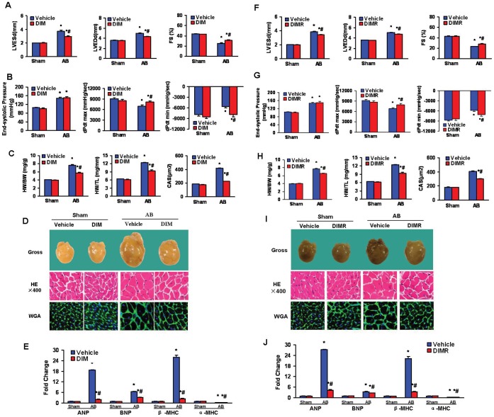
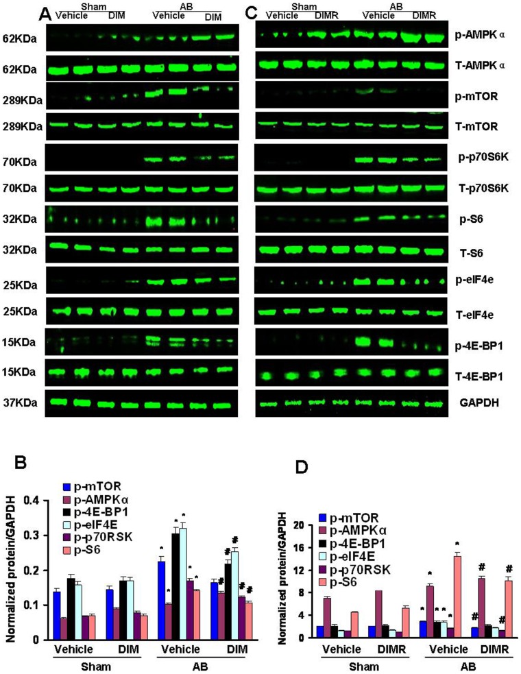
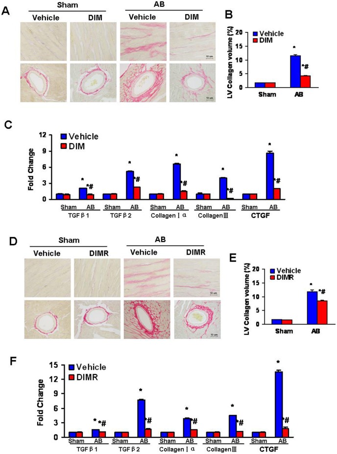
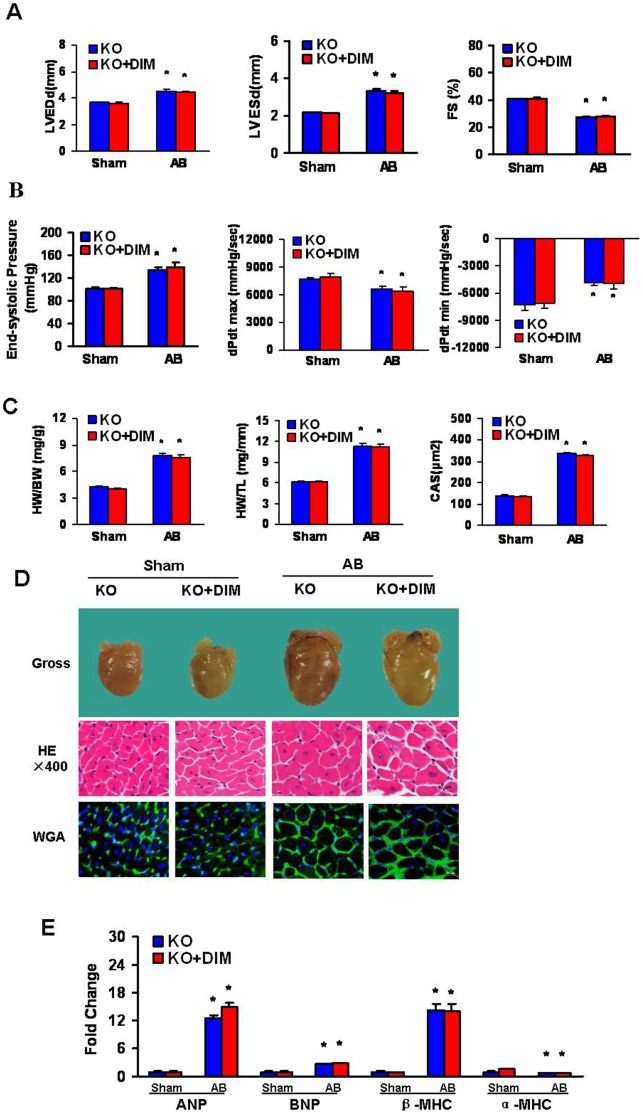
Results: In pre-treatment and reverse experiments, C57/BL6 mouse chow containing 0.05% DIM (dose 100 mg/kg/d DIM) was administered one week prior to surgery or one week after surgery, respectively, and continued for 8 weeks after surgery. In both experiments, DIM reduced to cardiac hypertrophy and fibrosis induced by aortic banding through the activation of 5'-adenosine monophosphate-activated protein kinase-α2 (AMPKα2) and inhibition of mammalian target of the rapamycin (mTOR) signaling pathway. Furthermore, DIM protected against cardiac oxidative stress by regulating expression of estrogen-related receptor-alpha (ERRα) and NRF2 etc. The cardioprotective effects of DIM were ablated in mice lacking functional AMPKα2.
Conclusion: DIM significantly improves left ventricular function via the activation of AMPKα2 in a murine model of cardiac hypertrophy.
Conflict of interest statement
Figures
Similar articles
-
3,3'-Diindolylmethane attenuates cardiac H9c2 cell hypertrophy through 5'-adenosine monophosphate-activated protein kinase-α.Mol Med Rep. 2015 Jul;12(1):1247-52. doi: 10.3892/mmr.2015.3523. Epub 2015 Mar 20. Mol Med Rep. 2015. PMID: 25816057
-
Indole-3-carbinol protects against pressure overload induced cardiac remodeling via activating AMPK-α.Mol Nutr Food Res. 2013 Sep;57(9):1680-7. doi: 10.1002/mnfr.201300012. Epub 2013 Apr 27. Mol Nutr Food Res. 2013. PMID: 23625645
-
Resistin promotes cardiac hypertrophy via the AMP-activated protein kinase/mammalian target of rapamycin (AMPK/mTOR) and c-Jun N-terminal kinase/insulin receptor substrate 1 (JNK/IRS1) pathways.J Biol Chem. 2011 May 27;286(21):18465-73. doi: 10.1074/jbc.M110.200022. Epub 2011 Apr 8. J Biol Chem. 2011. PMID: 21478152 Free PMC article.
-
Cordycepin ameliorates cardiac hypertrophy via activating the AMPKα pathway.J Cell Mol Med. 2019 Aug;23(8):5715-5727. doi: 10.1111/jcmm.14485. Epub 2019 Jun 21. J Cell Mol Med. 2019. PMID: 31225721 Free PMC article.
-
Regression of pathological cardiac hypertrophy: signaling pathways and therapeutic targets.Pharmacol Ther. 2012 Sep;135(3):337-54. doi: 10.1016/j.pharmthera.2012.06.006. Epub 2012 Jun 29. Pharmacol Ther. 2012. PMID: 22750195 Free PMC article. Review.
Cited by
-
Hesperetin protects against cardiac remodelling induced by pressure overload in mice.J Mol Histol. 2013 Oct;44(5):575-85. doi: 10.1007/s10735-013-9514-7. Epub 2013 May 30. J Mol Histol. 2013. PMID: 23719775
-
Characterization of RA839, a Noncovalent Small Molecule Binder to Keap1 and Selective Activator of Nrf2 Signaling.J Biol Chem. 2015 Nov 20;290(47):28446-28455. doi: 10.1074/jbc.M115.678136. Epub 2015 Oct 12. J Biol Chem. 2015. PMID: 26459563 Free PMC article.
-
New approaches to radiation protection.Front Oncol. 2015 Jan 20;4:381. doi: 10.3389/fonc.2014.00381. eCollection 2014. Front Oncol. 2015. PMID: 25653923 Free PMC article. Review.
-
Diindolylmethane Ameliorates Ischemic Stroke-Induced Brain Injury by Peripheral and Central Mechanisms.Curr Neurovasc Res. 2022;19(5):462-475. doi: 10.2174/1567202620666221116161128. Curr Neurovasc Res. 2022. PMID: 36397630
-
Protection against cardiac hypertrophy by geniposide involves the GLP-1 receptor / AMPKα signalling pathway.Br J Pharmacol. 2016 May;173(9):1502-16. doi: 10.1111/bph.13449. Epub 2016 Mar 14. Br J Pharmacol. 2016. PMID: 26845648 Free PMC article.
References
-
- Gupta S, Das B, Sen S (2007) Cardiac hypertrophy: mechanisms and therapeutic opportunities. Antioxid Redox Signal 9: 623–652. - PubMed
-
- Barry SP, Davidson SM, Townsend PA (2008) Molecular regulation of cardiac hypertrophy. Int J Biochem Cell Biol 40: 2023–2039. - PubMed
-
- Balakumar P, Singh AP, Singh M (2007) Rodent models of heart failure. J Pharmacol Toxicol Methods 56: 1–10. - PubMed
-
- Li Y, Wang Z, Kong D, Murthy S, Dou QP, et al. (2007) Regulation of FOXO3a/beta-catenin/GSK-3beta signaling by 3,3′-diindolylmethane contributes to inhibition of cell proliferation and induction of apoptosis in prostate cancer cells. J Biol Chem 282: 21542–21550. - PubMed
Publication types
MeSH terms
Substances
LinkOut - more resources
Full Text Sources
Other Literature Sources
Miscellaneous
