. 2013;8(1):e53491.
doi: 10.1371/journal.pone.0053491. Epub 2013 Jan 11.

Antitumor activity of antimicrobial peptides containing CisoDGRC in CD13 negative breast cancer cells

Affiliations

Antitumor activity of antimicrobial peptides containing CisoDGRC in CD13 negative breast cancer cells

Lei Hou et al. PLoS One. 2013.

Abstract

Background: isoAsp-Gly-Arg (isoDGR) is a derivative of the Asn-Gly-Arg (NGR) motif, which is used as a targeted delivery tool to aminopeptidase N (CD13) positive cells. Recent studies have shown that cyclic isoDGR (CisoDGRC) has a more efficient affinity with α(v)β(3), a type of integrin that overexpresses in tumor cells. Antimicrobial peptides (AMPs) are an efficient antitumor peptide that specifically kills tumor cells. In the present study, we designed antimicrobial peptides containing the CisoDGRC motif (CDAK) and assessed its antitumor activity for CD13(-)/α(v)β(3) (+) breast cancer cells (MCF-7 and MDA-MB-231) in vitro and in vivo.

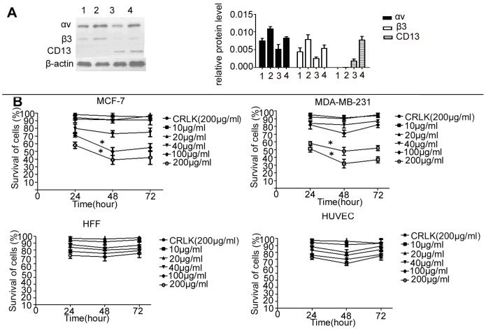
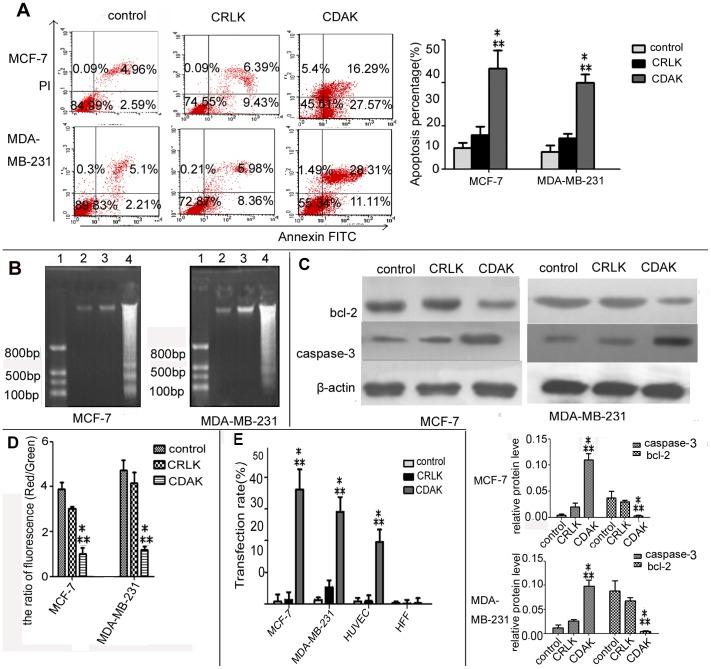
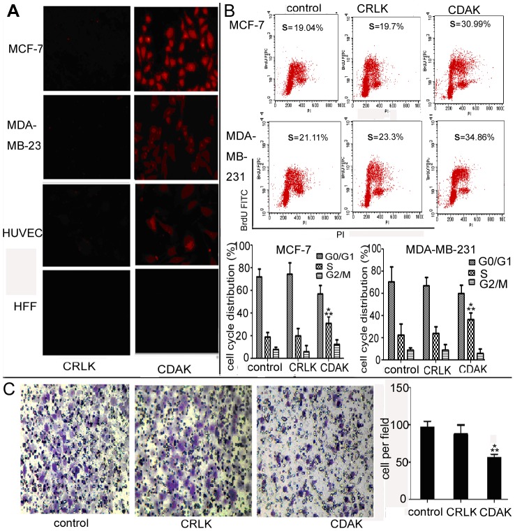
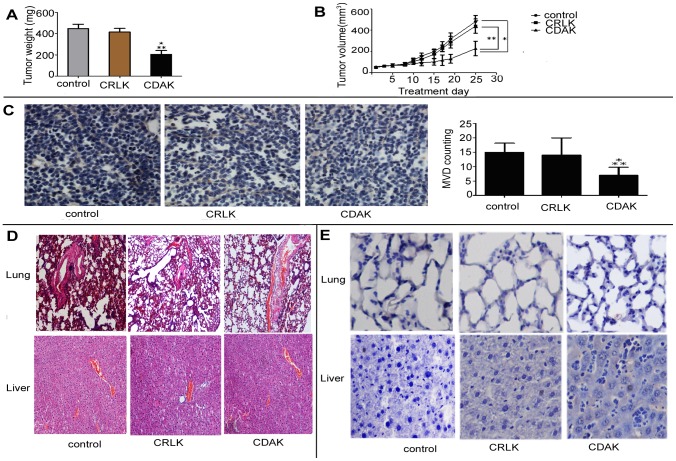
In vitro: We assessed the cytotoxicity of CDAK for MCF-7 and MDA-MB-231 breast cancer cells, the human umbilical vein endothelial cell (HUVEC), and human foreskin fibroblasts (HFF). We performed an apoptosis assay using Annexin-V/PI, DNA ladder, mitochondrial membrane potential, and Caspase-3 and Bcl-2. The effect on cell cycles and affinity with cell were tested using flow cytometry and fluorescent microscopy and the effect on invasion was analyzed using an invasion assay. CDAK was injected intravenously into tumor-bearing athymic nude mice in vivo experiment.

Results: CDAK showed cytotoxic activity in MCF-7 and MDA-MB-231 cells, whereas HUVEC and HFF were less sensitive to the peptides. CDAK induced apoptosis, reduced mitochondrial membrane potential, promoted Caspase-3, and inhibited Bcl-2 expression in the two breast cancer cell lines. In addition, CDAK inhibited proliferation of cancer cell through S phase arrest, and own selective affinity with MCF-7 and MDA-MB-231cells, inhibited the invasion of MDA-MB-231 cells. In vivo, CDAK significant inhibited the progression of the tumor and the generation of neovascularization.

Conclusion: Antimicrobial peptides containing the CisoDGRC (CDAK) motif could efficiently exhibit the antitumor activity for CD13(-)/α(v)β(3) (+) breast cancer cells.


Conflict of interest statement

Competing Interests: The authors have declared that no competing interests exist.

Figures

Figure 1
Figure 1. The expression status of αvβ3 and CD13 and the cytotoxic activity of CDAK in cell lines.
(A) Western-blot analyzed the protein expression of αvβ3 and CD13 in the four cell lines, Lane 1–4, MDA-MB-231, MCF-7, HFF and HUVEC. The expression levels were analyzed by the ratio of optical density with β-actin. P = 0.005 (ANOVA asay) (B) The four cell lines were cultured with CRLK (200 µg/ml) and various concentrations of CDAK (10–200 µg/ml) for 24 h, 48 h, and 72 h. The cytotoxic activity was assessed using MTT. CDAK had significant cytotoxicity for MCF-7 and MDA-MB-231cells, P<0.01 (ANOVA asay). Data are presented by means ± SD (bar) from triplicate determinations. *P<0.05 versus control.
Figure 2
Figure 2. Characterization of the breast cancer cell death mechanism by CDAK.
(A) The apoptosis of breast cancer cells treated with CRLK (200 µg/ml) and CDAK (190 µg/ml in MCF-7, 212 µg/ml in MDA-MB-231) for 24 h were analyzed using flow cytometry for Annexin V/PI stain. The sum of the upper right and lower right quadrants are expressed as a percentage of the apoptotic cell. CDAK enhanced the percentage ratio of apoptosis, P<0.01. (B) Electrophoretic analysis of DNA extracted from MCF-7 and MDA-MB-231 treated with CRLK(200 µg/ml) and CDAK(190 µg/ml,212 µg/ml) for 24 h. Lane 1, DNA mark; Lane 2–4, control, CRLK and CDAK. The CDAK group forms a clear DNA ladder. (C) Pro-apoptosis protein caspase-3 and inhibit protein bcl-2 were analyzed by Western-blot. The whole protein of MCF-7 and MDA-MB-231 treated with CRLK (200 µg/ml) and CDAK (190 µg/ml,212 µg/ml) for 24 h were extracted. The expression levels were analyzed by the ratio of optical density with β-actin. CDAK inhibited the expression of bcl-2 promoted caspase-3 compared with CRLK and control, P<0.01. (D) MCF-7 and MDA-MB-231 treated with CRLK (200 µg/ml) and CDAK (190 µg/ml and 212 µg/ml) for 24 h were analyzed for the mitochondrial transmembrane potential using flow cytometry and JC-1. The ratio of fluorescence (red/green) indicates the mitochondrial transmembrane potential. CDAK decreased the ration of fluorescence, P<0.01. (E) The affinity rate of CRLK and CDAK (10 µg/ml) for 12 h were analyzed using flow cytometry. MCF-7 and MDA-MB-231 showed more binding than HUVEC and HFF, especially MCF-7, P<0.01 (ANOVA assay). The results are represented as means± SD from triplicate determinations. * P<0.05 versus control; ** P<0.05 versus CRLK.
Figure 3
Figure 3. The affinity of CDAK and the effect of CDAK on cell cycle and invasion.
(A) Fluorescence photograph of MCF-7, MDA-MB-231, HUVEC and HFF cells treated with CRLK and CDAK (10 µg/ml), which is labeled by Rhodamine B in the K of the amino acid residues' end, for 12 h (400× magnification). (B) MCF-7 and MDA-MB-231 cells treated with CRLK (200 µg/ml) and CDAK (190 µg/ml in MCF-7 and 212 µg/ml in MDA-MB-231) for 24 h were analyzed for the cell cycle distribution using flow cytometry for BrdU/PI stain. CDAK induced the S phage arrest, P<0.01 (ANOVA asay). (C) Representative image depicting the effect of CDAK and CRLK (10 µg/ml) on MDA-MB-231 cells invasion (400× magnification). Quantitation of migrating cells per high field, P<0.01 (ANOVA assay). The data are mean ± SD from triplicate determinations. * P<0.05 versus control ** P<0.05 versus CRLK.
Figure 4
Figure 4. Antitumor activity of CDAK in breast cancer xenograft in vivo.
(A) Tumor–growth curve. MDA-MB-231 cell lines were implanted into the right flank of female athymic mice. When the tumor size reached about 60 mm3, mice received an intravenous injection of CDAK (4 mg/kg,) and CRLK (4 mg/kg), or saline used as control (three times a week). The volumes of the tumors were measure three times a week. The tumor volume of CDAK group was smaller than control and CRLK, P<0.01. (B) The athymic mice were sacrificed as 25 d after being injected with CDAK and CRLK and the weight of each tumor was measured. The CDAK group had low weight compared to the control and the CRLK groups. P = 0.003. (C) The inhibition of angiogenesis was evaluated in tumor sections using immunohistochemistry with anti-CD105 antibody. Angiogenesis was quantified by image analysis of Microvessel density (MVD) (400× magnification). CDAK caused significant inhibition of angiogenesis. P = 0.003. (D) Images (200× magnification) of the lung and liver from athymic mice were obtained by staining with hemaetoxylin and eosin (HE). No significant damage was observed. (E) Representative photographs of the tumor section examined by TUNEL assay, light microscope (×400). The number of apoptotic cells was counter 5 random field in blinded manner. There differences of apoptotic cell were not statistic significant in the three groups (table 2) (ANOVA assay). The images were analyzed by pro plus 5.0. Each group had five nude mice. Data are shown as mean ± SD. *P<0.05 versus control, ** P<0.05 versus CRLK.

Similar articles

Cited by

References

    1. Zhou T, Jia X, Li H, Wang J, Zhang H, et al. (2011) New tumor-targeted nanosized delivery carrier for oligonucleotides: characteristics in vitro and in vivo. Int J Nanomedicine 6: 1527–1534. - PMC - PubMed
    1. Gregorc V, De Braud FG, De Pas TM, Scalamogna R, Citterio G, et al. (2011) Phase I study of NGR-hTNF, a selective vascular targeting agent, in combination with cisplatin in refractory solid tumors. Clin Cancer Res 17: 1964–1972. - PubMed
    1. Chen Y, Wu JJ, Huang L (2010) Nanoparticles targeted with NGR motif deliver c-myc siRNA and doxorubicin for anticancer therapy. Mol Ther 18: 828–834. - PMC - PubMed
    1. Rizzardi GP, Bordignon C (2009) NGR and isoDGR are separate moieties binding to different receptors. Blood 113: 5366; author reply 5367. - PubMed
    1. Corti A, Curnis F (2011) Isoaspartate-dependent molecular switches for integrin-ligand recognition. J Cell Sci 124: 515–522. - PubMed

Publication types

MeSH terms