. 2013;8(1):e53564.
doi: 10.1371/journal.pone.0053564. Epub 2013 Jan 9.

Tumor-derived autophagosomes (DRibbles) induce B cell activation in a TLR2-MyD88 dependent manner

Affiliations

Tumor-derived autophagosomes (DRibbles) induce B cell activation in a TLR2-MyD88 dependent manner

Weixia Li et al. PLoS One. 2013.

Abstract

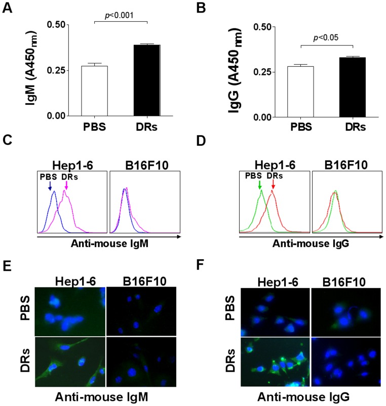
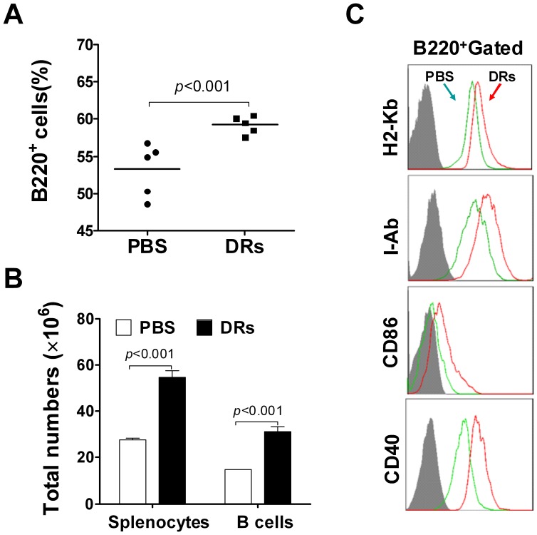
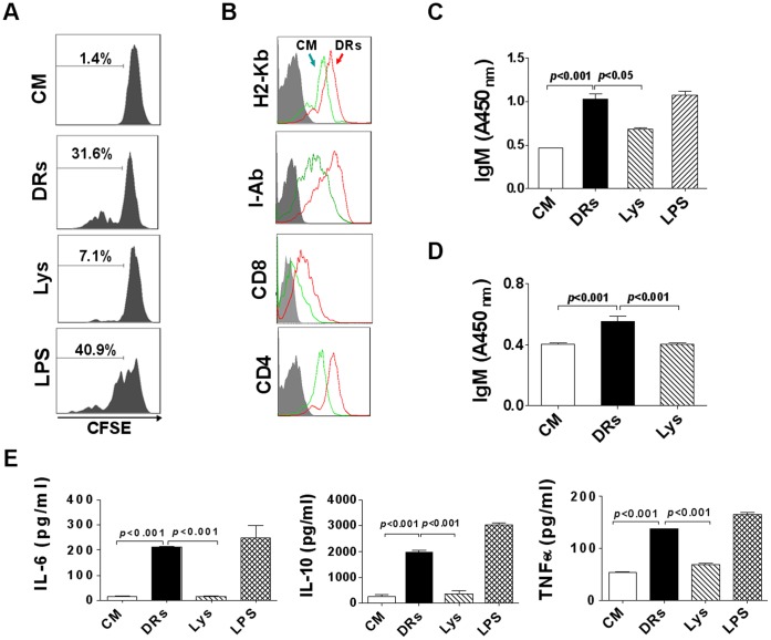
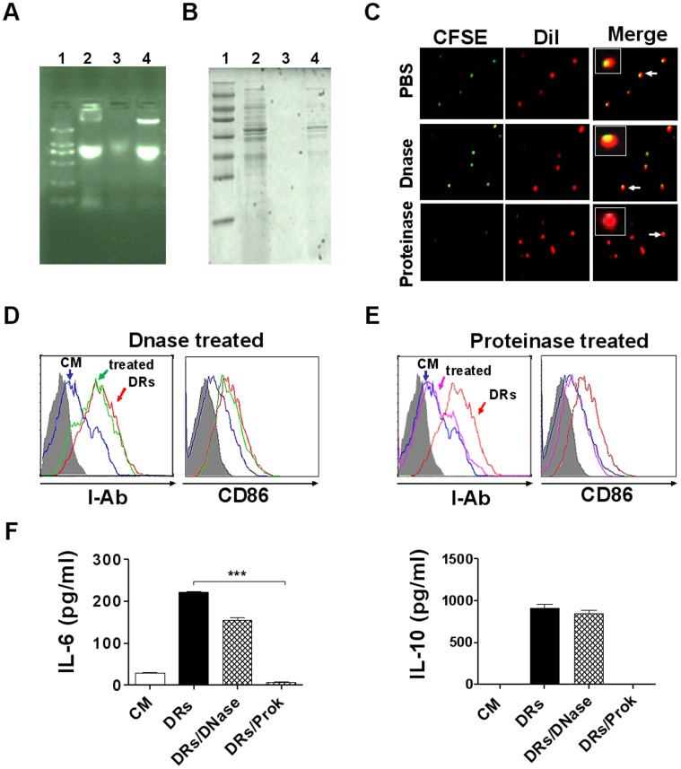
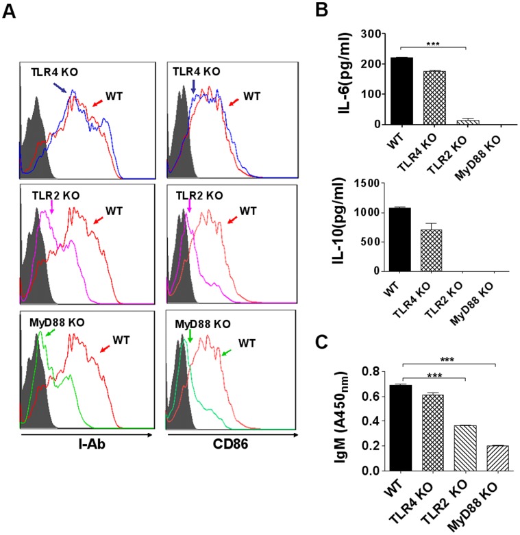
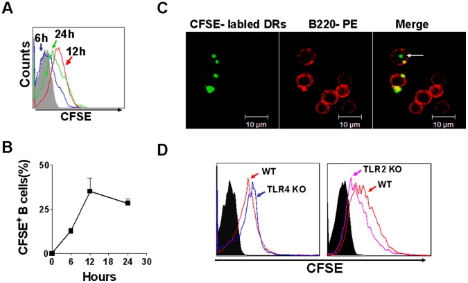
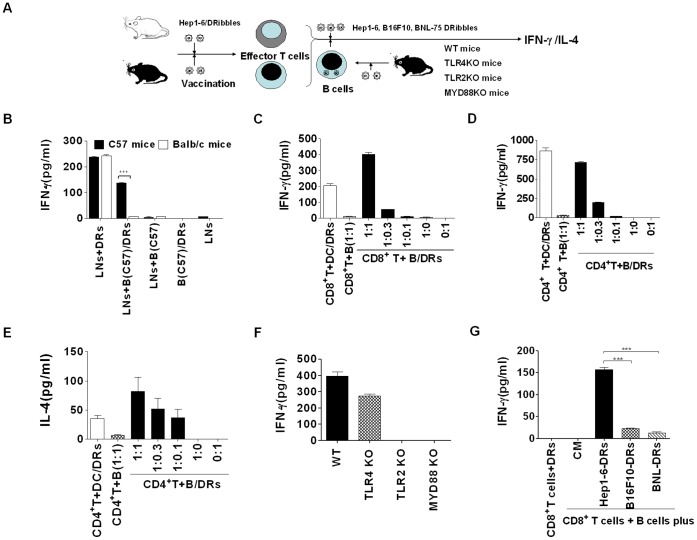
Previously, we have documented that isolated autophagosomes from tumor cells could efficiently cross-prime tumor-reactive naïve T cells and mediate tumor regression in preclinical mouse models. However, the effect of tumor-derived autophagosomes, here we refer as to DRibbles, on B cells has not been studied so far. At present study, we found that DRibbles generated from a murine hepatoma cell line Hep1-6, induced B-cell activation after intravenous injection into mice. B-cell populations were significantly expanded and the production of Hep1-6 tumor-specific antibodies was successfully induced. Moreover, in vitro studies showed that DRibbles could induce more efficient B-cell proliferation and activation, antibody production, and cytokine secretion than whole tumor cell lysates. Notably, we found that B-cell activation required proteins but not DNA in the DRibbles. We further showed that B cells could capture DRibbles and present antigens in the DRibbles to directly induce T cell activation. Furthermore, we found that B-cell activation, antibody production, cytokine secretion and antigen cross-presentation were TLR2-MyD88 pathway dependent. Taken together, the present studies demonstrated that tumor-derived autophagosomes (DRibbles) efficiently induced B cells activation, antibody production, cytokine secretion and antigen cross-presentation mainly depending on their protein component via TLR2/MyD88 dependent manner.


Conflict of interest statement

Competing Interests: The authors have declared that no competing interests exist.

Figures

Figure 1
Figure 1. DRibbles derived from tumor cells induce specific antibody production in vivo.
(A and B) Serum was collected from C57/BL6 mice at day 7 after first intravenous injection of PBS or Hep1-6 derived DRibbles (DRs). The total IgM (A) and IgG (B) in serum was measured by ELISA. Values are the mean ± SEM derived from three mice per group (n = 3). (C and D) Hep1-6 or B16F10 cells were incubated for 1 hour with serum derived from PBS or Hep1-6 DRibbles injected mice respectively. After washing, Hep1-6 or B16F10 cells were incubated with FITC-labeled anti-mouse IgM antibody (C) or FITC-labeled anti-mouse IgG antibody (D). After washing, the cells were analyzed by flow cytometry. (E and F) HepI-6 or B16F10 cells were treated as C and D, and then the cells were stained with DAPI and analyzed by fluorescence microscope. Green fluorescence represents FITC-labeled anti-mouse IgM antibody (E) or FITC-labeled anti-mouse IgG antibody (F). Blue fluorescence represents the cell nucleus. A representative of three independent experiments was showed.
Figure 2
Figure 2. DRibbles induced proliferation and activation of B cells in vivo.
(A and B) C57/BL6 mice were intravenously injected with DRibbles (DRs) or PBS as the control for 3 times in day 1, 2 and 3. Splenocytes were collected from each mouse in day 4 after first injection and were stained with anti-mouse B220 mAb. The percentage of B220+ cells in splenocytes (A) and total number of splenocytes and B220+ cells from each mouse (B) were analyzed by flow cytometry (n = 5). (C) Splenocytes from each mouse were stained with anti-mouse B220 mAb and the indicated mAbs respectively. The expression of H2-Kb, I-Ab, CD40 and CD86 of B220+ gated cells was analyzed by flow cytometry. Gray shaded histograms indicate the isotype staining. Results represent three independent experiments.
Figure 3
Figure 3. DRibbles induced proliferation and activation of B cells in vitro.
(A) Purified B cells were labeled with CFSE and then co-incubated with DRibbles (DRs), whole tumor cell lysate (Lys) or LPS for 5 days. The proliferation of B cells was accessed by flow cytometry. (B) Purified B cells were co-incubated with DRibbles for 3 days and collected for staining with the indicated Abs or isotype-matched control Abs (gray filled area). The expression of H2-Kb, I-Ab, CD86 and CD40 on B cells was analyzed by flow cytometry. (C and D) Splenocytes (C) and purified B cells (D) were co-incubated with DRibbles or lysate respectively for 7 days. IgM in the supernatants was analyzed by ELISA. (E) Purified B cells were co-incubated with DRibbles, tumor cell lysate or LPS for 3 days. Cytokines including IL-6, IL-10, TNF-αα in the supernatants was analyzed by ELISA. (CM indicated complete medium). Results represent three independent experiments.
Figure 4
Figure 4. B cells activation was mainly depended on proteins but not DNA in DRibbles.
DRibbles (DRs) were digested with DNase I or proteinase K and then released after lysed with RIPA lysis buffer. (A) The DNA in DRibbles was analyzed via agarose gel electrophoresis. (1. Markers 2. DRibbles 3. DRibbles digested with DNase I, 4. DRibbles digested with proteinase K). (B) The proteins in DRibbles were analyzed via 12% SDS polyacrylamide gel electrophoresis. (1. Markers 2. DRibbles 3. DRibbles digested with Proteinase K 4. DRibbles digested with DNase I). (C) DRibbles, DRibbles digested with DNase I or Proteinase K were stained with both CFSE and DiI. The characteristic of DRibbles was detected by the fluorescent microscope analysis (green presents CFSE labeled DRibbles-proteins; red presents DiI labeled DRibbles-membrane). (D and E) DRibbles digested with or without DNase I (D) or proteinase K (E) were co-incubated with purified B cells for 72 hours. B cells were collected and stained with Abs to corresponding surface markers (indicated), or isotype-matched control Ab (gray filled area). The expression of I-Ab and CD86 on B cells were analyzed by flow cytometry (blue line indicated complete medium; red line indicated DRibbles; green or pink line indicated DRibbles treated with DNase or proteinase). (F) B cells were incubated with DRibbles digested with or without DNase I or proteinase K for 72 hours, IL-6 and IL-10 in the supernatants were detected by ELISA. Results represent three independent experiments.
Figure 5
Figure 5. The role of TLR4, TLR2 and MyD88 signaling pathways in B cells activation induced by DRibbles.
B cells purified from wild type, TLR4-, TLR2- or MyD88-deficient mice were co-incubated with DRibbles respectively for 72 hours. (A) B cells was collected and stained with indicated Abs. The expression of I-Ab and CD86 on B cells were detected by flow cytometer. (B) IL-6 and IL-10 in the supernatants were determined by ELISA. (C) IgM in the supernatants was determined by ELISA. (WT indicated B cells from wild type mice; TLR4KO, TLR2KO, and MyD88KO indicated B cells from TLR4-, TLR2- and MyD88-deficient mice). Results represent three independent experiments.
Figure 6
Figure 6. DRibbles can be taken up by B cells in vitro.
(A) Purified B cells were co-incubated with CFSE-labeled DRibbles for 6, 12, 24 hours, CFSE positive B cells were assessed by flow cytometry. (B) The percentage of CFSE positive B cells were counted by flow cytometry as it has been indicated. (C) B cells taking up DRibbles were detected by the confocal laser microscope analysis. Representative images of immunofluorescence were stained with PE-conjugated anti-mouse B220 (red) and DRibbles labeled with CFSE (green). Arrows indicated that CFSE-labeled DRibbles were located inside of B cells. (D) B cells purified from wild type, TLR4- and TLR2-deficient mice were co-incubated with CFSE labeled-DRibbles for 12 hours, then the CFSE positive B cells were analyzed by flow cytometer. Data represent one of at least three experiments with similar results.
Figure 7
Figure 7. B cells loading DRibbles antigen could re-activate effector T cells.
(A) The schematic diagram outlines the experiment protocol. C57/BL6 mice or BALB/c mice were intro-node vaccinated with DRibbles respectively. Lymphocytes were collected from lymph nodes (LN) of vaccinated C57/BL6 mice or BALB/c mice on day 7 after immunization. B cells purified from wide type C57/BL6 mice were stimulated with DRibbles for 6 hours and then washed 3 times with PBS. The lymphocytes were co-incubated with DRibbles or DRibbles-loaded B cells or B cells alone for 72 hours (n = 5). (B) The supernatants were harvested for detection of IFN-γ by ELISA. (C) CD8+ T cells were purified from the vaccinated C57/BL6 mice, and then co-incubated with B cells plus DRibbles (with DCs plus DRibbles at 1∶1 ratio or B cell alone as control) at the indicated ratio for 72 hours. IFN-γ in the supernatants was tested via ELISA. (D and E) CD4+ T cells were purified from the vaccinated C57/BL6 mice, and then co-incubated with B cells plus DRibbles at the indicated ratio for 72 hours. IFN-γ (D) and IL-4 (E) in the supernatants were tested via ELISA. (F) CD8+ T cells purified from the DRibbles-vaccinated C57/BL6 mice were co-incubated at 1∶1 ratio with B cells (from wild type, TLR4-, TLR2- or MyD88-deficient mice) plus DRibbles for 72 hours. IFN-γ in the supernatants was tested via ELISA. (G) CD8+ T cells were purified from the Hep1-6-DRibbles vaccinated C57/BL6 mice, and then co-incubated at 1∶1 ratio with B cells plus Hep1-6-, B16F10- or BNL-DRibbles for 72 hours respectively. IFN-γ in the supernatants was tested via ELISA. Data which are presented were obtained as a result of triplicates.

Similar articles

Cited by

References

    1. Abeliovich H, Dunn WAJ, Kim J, Klionsky DJ (2000) Dissection of autophagosome biogenesis into distinct nucleation and expansion steps. J Cell Biol 151: 1025–1034. - PMC - PubMed
    1. Li YH, Wang LX, Pang P, Twitty C, Fox BA, et al. (2009) Cross presentation of tumor associated antigens through tumor-derived autophagosomes. Autophagy 5: 576–577. - PMC - PubMed
    1. Li Y, Wang LX, Yang G, Hao F, Urba WJ, et al. (2008) Efficient Cross-presentation Depends on Autophagy in Tumor Cells. Cancer Res 68: 6889–6895. - PMC - PubMed
    1. Li YH, Wang LX, Pang P, Cui Z, Aung S, et al. (2011) Tumor-derived autophagosome vaccine: mechanism of cross-presentation and therapeutic efficacy. Clin Cancer Res 17(00): 1–11. - PMC - PubMed
    1. Twitty CG, Jensen SM, Hu HM, Fox BA (2011) Tumor-derived autophagosome vaccine: induction of cross-protective immune responses against short-lived proteins through a p62-dependent mechanism. Clin Cancer Res 17: 6467–6481. - PMC - PubMed

Publication types

MeSH terms