

Construction and accuracy assessment of patient-specific biocompatible drill template for cervical anterior transpedicular screw (ATPS) insertion: an in vitro study
- PMID: 23326461
- PMCID: PMC3542371
- DOI: 10.1371/journal.pone.0053580
Construction and accuracy assessment of patient-specific biocompatible drill template for cervical anterior transpedicular screw (ATPS) insertion: an in vitro study
Abstract
Background: With the properties of three-column fixation and anterior-approach-only procedure, anterior transpedicular screw (ATPS) is ideal for severe multilevel traumatic cervical instabilities. However, the accurate insertion of ATPS remains challenging. Here we constructed a patient-specific biocompatible drill template and evaluated its accuracy in assisting ATPS insertion.
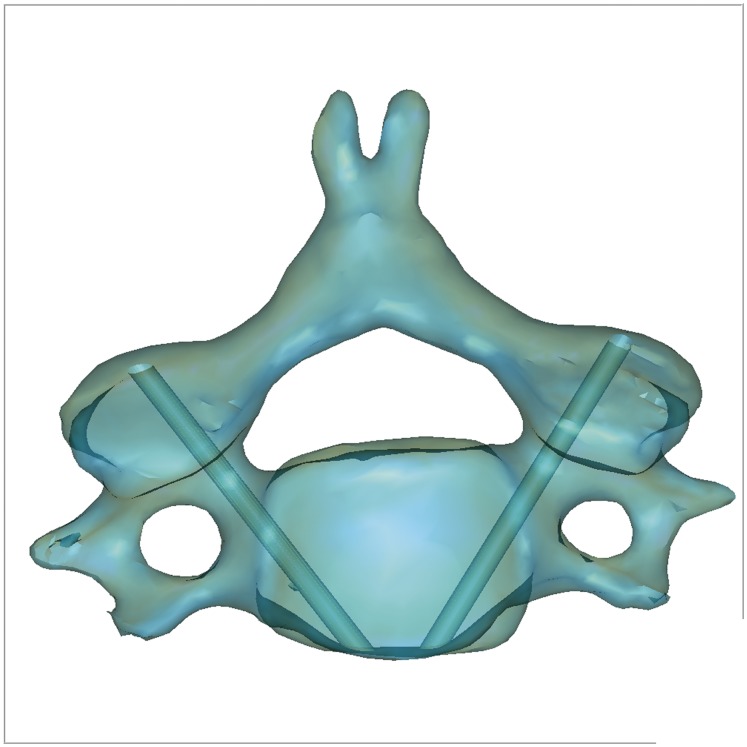
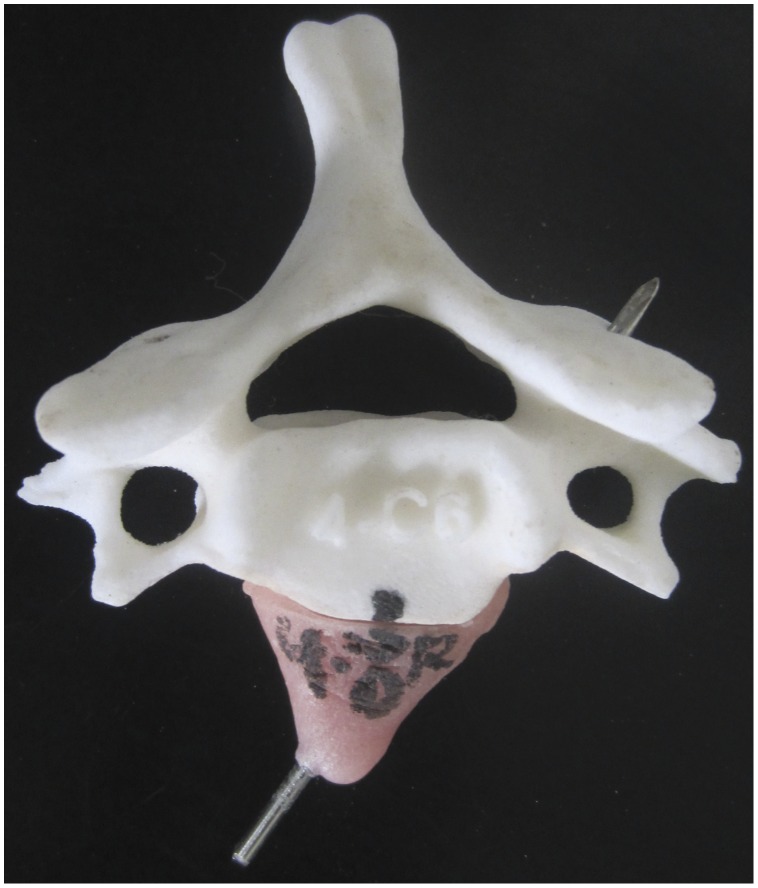
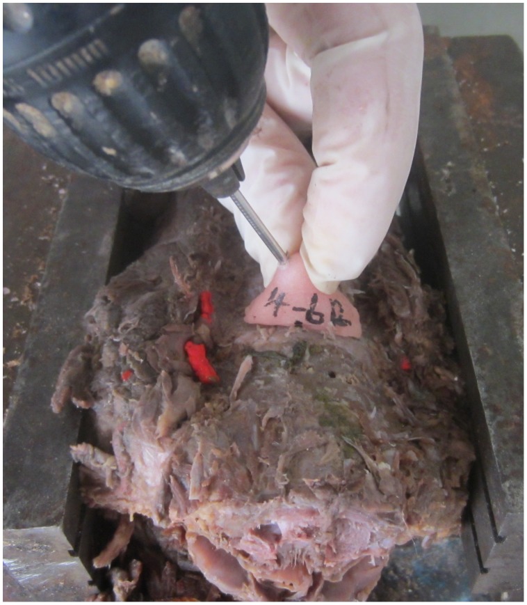
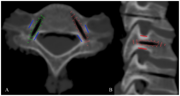
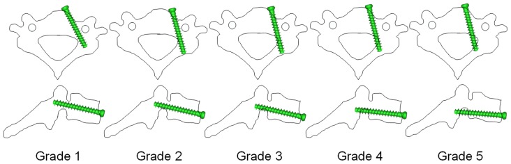
Methods: After ethical approval, 24 formalin-preserved cervical vertebrae (C2-C7) were CT scanned. 3D reconstruction models of cervical vertebra were obtained with 2-mm-diameter virtual pin tracts at the central pedicles. The 3D models were used for rapid prototyping (RP) printing. A 2-mm-diameter Kirschner wire was then inserted into the pin tract of the RP model before polymethylmethacrylate was used to construct the patient-specific biocompatible drill template. After removal of the anterior soft tissue, a 2-mm-diameter Kirschner wire was inserted into the cervical pedicle with the assistance of drill template. Cadaveric cervical spines with pin tracts were subsequently scanned using the same CT scanner. A 3D reconstruction was performed of the scanned spines to get 3D models of the vertebrae containing the actual pin tracts. The deviations were calculated between 3D models with virtual and actual pin tracts at the middle point of the cervical pedicle. 3D models of 3.5 mm-diameter screws were used in simulated insertion to grade the screw positions.
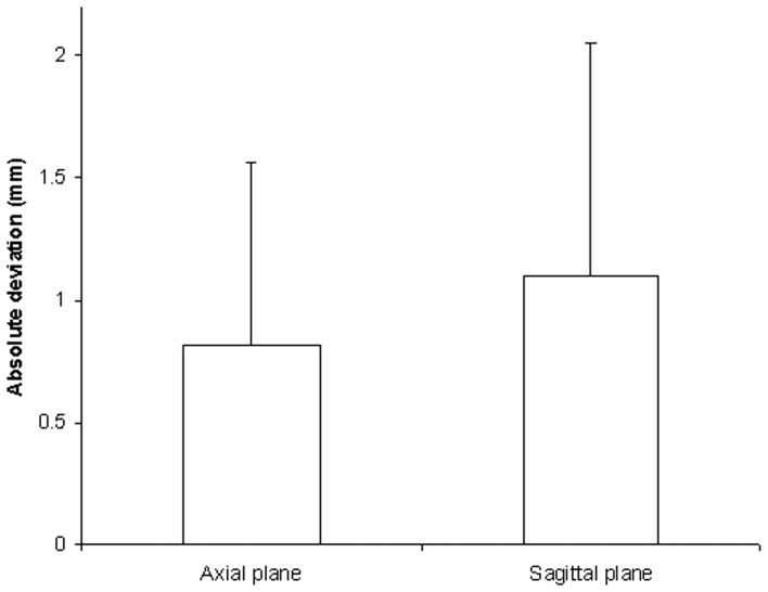
Findings: The patient-specific biocompatible drill template was constructed to assist ATPS insertion successfully. There were no significant differences between medial/lateral deviations (P = 0.797) or between superior/inferior deviations (P = 0.741). The absolute deviation values were 0.82±0.75 mm and 1.10±0.96 mm in axial and sagittal planes, respectively. In the simulated insertion, the screws in non-critical position were 44/48 (91.7%).
Conclusions: The patient-specific drill template is biocompatible, easy-to-apply and accurate in assisting ATPS insertion. Its clinical applications should be further researched.
Conflict of interest statement
Figures
Similar articles
-
Is a patient-specific drill template via a cortical bone trajectory safe in cervical anterior transpedicular insertion?J Orthop Surg Res. 2018 Apr 18;13(1):91. doi: 10.1186/s13018-018-0810-5. J Orthop Surg Res. 2018. PMID: 29669577 Free PMC article.
-
Cervical anterior transpedicular screw fixation (ATPS)--Part II. Accuracy of manual insertion and pull-out strength of ATPS.Eur Spine J. 2008 Apr;17(4):539-55. doi: 10.1007/s00586-007-0573-x. Epub 2008 Jan 26. Eur Spine J. 2008. PMID: 18224357 Free PMC article.
-
A novel patient-specific navigational template for cervical pedicle screw placement.Spine (Phila Pa 1976). 2009 Dec 15;34(26):E959-66. doi: 10.1097/BRS.0b013e3181c09985. Spine (Phila Pa 1976). 2009. PMID: 20010385
-
Anterior transpedicular screw fixation of cervical spine: Is it safe? Morphological feasibility, technical properties, and accuracy of manual insertion.J Neurosurg Spine. 2015 Jun;22(6):596-604. doi: 10.3171/2014.10.SPINE14669. Epub 2015 Mar 27. J Neurosurg Spine. 2015. PMID: 25815805
-
A novel computer-assisted drill guide template for lumbar pedicle screw placement: a cadaveric and clinical study.Int J Med Robot. 2009 Jun;5(2):184-91. doi: 10.1002/rcs.249. Int J Med Robot. 2009. PMID: 19280584
Cited by
-
Application of a customized 3D-printed osteotomy guide plate for tibial transverse transport.Sci Rep. 2024 Oct 1;14(1):22771. doi: 10.1038/s41598-024-73715-y. Sci Rep. 2024. PMID: 39354073 Free PMC article.
-
Patient-specific drill template for C2 transoral pedicle insertion in complete reduction of atlantoaxial dislocation: cadaveric efficacy and accuracy assessments.J Orthop Surg Res. 2019 May 16;14(1):141. doi: 10.1186/s13018-019-1189-7. J Orthop Surg Res. 2019. PMID: 31096990 Free PMC article. Clinical Trial.
-
Is a patient-specific drill template via a cortical bone trajectory safe in cervical anterior transpedicular insertion?J Orthop Surg Res. 2018 Apr 18;13(1):91. doi: 10.1186/s13018-018-0810-5. J Orthop Surg Res. 2018. PMID: 29669577 Free PMC article.
-
Error Analysis: How Precise is Fused Deposition Modeling in Fabrication of Bone Models in Comparison to the Parent Bones?Indian J Orthop. 2018 Mar-Apr;52(2):196-201. doi: 10.4103/ortho.IJOrtho_312_16. Indian J Orthop. 2018. PMID: 29576649 Free PMC article.
-
Are computer numerical control (CNC)-manufactured patient-specific metal templates available for posterior thoracic pedicle screw insertion? Feasibility and accuracy evaluation.Eur Spine J. 2017 Nov;26(11):2927-2933. doi: 10.1007/s00586-017-5215-3. Epub 2017 Jul 17. Eur Spine J. 2017. PMID: 28718167
References
-
- Ponnusamy KE, Iyer S, Gupta G, Khanna AJ (2011) Instrumentation of the osteoporotic spine: biomechanical and clinical considerations. Spine J 11(1): 54–63. - PubMed
-
- Bozkus H, Ames CP, Chamberlain RH, Nottmeier EW, Sonntag VK, et al. (2005) Biomechanical analysis of rigid stabilization techniques for three-column injury in the lower cervical spine. Spine (Phila Pa 1976) 30(8): 915–922. - PubMed
Publication types
MeSH terms
Substances
LinkOut - more resources
Full Text Sources
Other Literature Sources
Medical
Miscellaneous
