. 2013;8(1):e53665.
doi: 10.1371/journal.pone.0053665. Epub 2013 Jan 9.

Genetic and functional dissection of ARMS2 in age-related macular degeneration and polypoidal choroidal vasculopathy

Affiliations

Genetic and functional dissection of ARMS2 in age-related macular degeneration and polypoidal choroidal vasculopathy

Yong Cheng et al. PLoS One. 2013.

Abstract

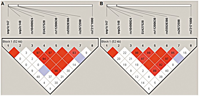
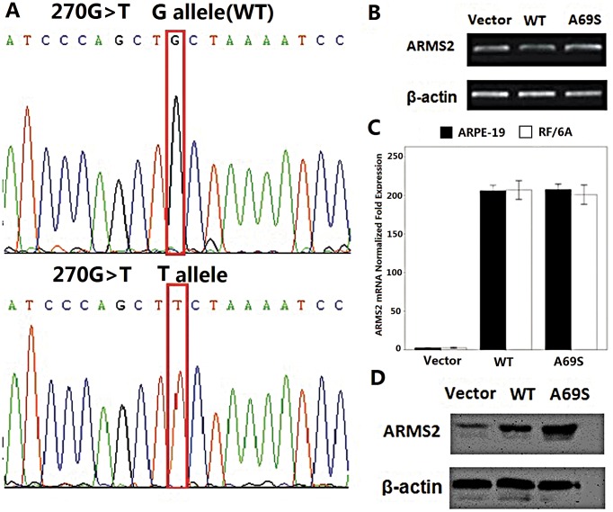
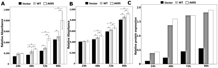
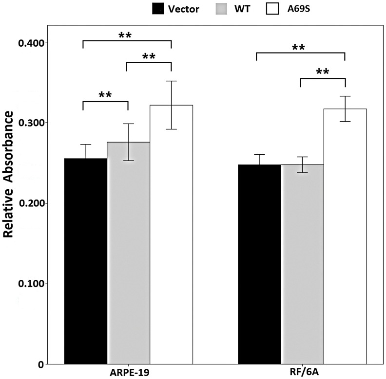
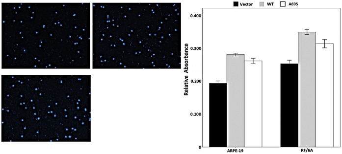
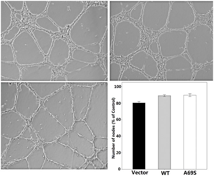
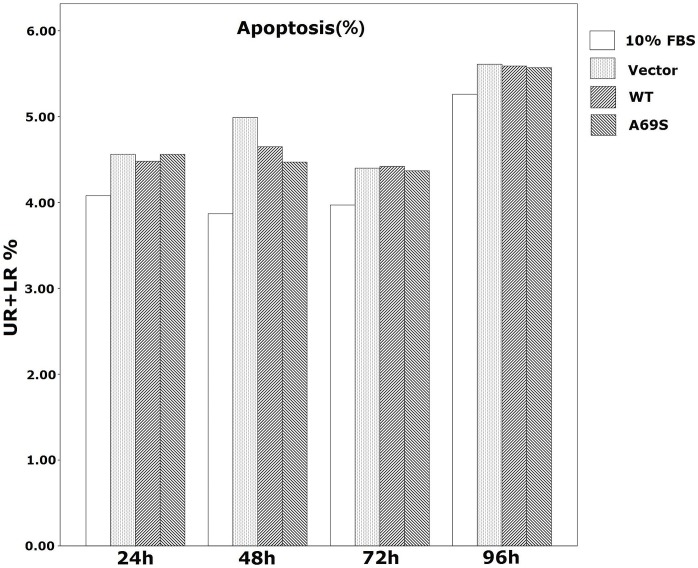
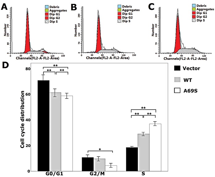
Age-related maculopathy susceptibility 2(ARMS2) was suggested to be associated with neovascular age-related macular degeneration (nAMD) and polypoidal choroidal vasculopathy (PCV) in multiple genetic studies in Caucasians and Japanese. To date, no biological properties have been attributed to the putative protein in nAMD and PCV. The complete genes of ARMS2 and HTRA1 including all exons and the promoter region were assessed using direct sequencing technology in 284 unrelated mainland northern Chinese individuals: 96 nAMD patients, 92 PCV patients and 96 controls. Significant associations with both nAMD and PCV were observed in 2 polymorphisms of ARMS2 and HTRA1 rs11200638, with different genotypic distributions between nAMD and PCV (p<0.001). After adjusting for rs11200638, ARMS2 rs10490924 remained significantly associated with nAMD and PCV (p<0.001). Then we overexpressed wild-type ARMS2 and ARMS2 A69S mutation (rs10490924) in RF/6A cells and RPE cells as in vitro study model. Cell proliferation, attachment, migration and tube formation were analyzed for the first time. Compare with wild-type ARMS2, A69S mutation resulted in a significant increase in proliferation and attachment but inhibited cell migration. Moreover, neither wild-type ARMS2 nor A69S mutation affected tube formation of RF/6A cells. There is a strong and consistent association of the ARMS2/HTRA1 locus with both nAMD and PCV, suggesting the two disorders share, at least partially, similar molecular mechanisms. Neither wild-type ARMS2 nor A69S mutation had direct association with neovascularisation in the pathogenesis of AMD.


Conflict of interest statement

Competing Interests: The authors have declared that no competing interests exist.

Figures

Figure 1
Figure 1. Analysis of pair-wise LD across ARMS2 and HTRA1 SNPs in northern Chinese PCV and nAMD cohort.
A) Analysis of pair-wise LD across the eight ARMS2 and HTRA1 SNPs in northern Chinese PCV cohort. B) Analysis of pair-wise LD across the eight ARMS2 and HTRA1 SNPs in northern Chinese nAMD cohort. The relative physical position of each SNP is given in the upper diagram. The pairwise D′ between all SNPs is given below each SNP combination. And when D′ = 1.0, no number is given inside the square. Bright red squares indicate D′≥0.90 and LOD≥2. Bright red squares indicate D′<0.90 and LOD≥2. White squares indicate D′<0.90 and LOD<2.
Figure 2
Figure 2. Rs10490924 (A69S in a coding change) does not affect mRNA, protein, or surface expression of ARMS2.
A)Direct sequencing verified the wild-type (WT) and G270T ARMS2 plasmids. B) RT-PCR showed no significant difference between wild-type and G270T ARMS2 mRNA expression. C) Real-time RT-PCR confirmed the mRNA level finding. D) Western blot showed that the levels and migrations of the SNP mutant proteins were comparable with wild type. All experiments were repeated >3 times.
Figure 3
Figure 3. Effect of wild-type ARMS2 and rs10490924 on the proliferation of RF/6A and human RPE cells.
RF/6A (A) and ARPE-19 (B) cell proliferation was measured with an MTT assay at 24 h, 48 h, 72 h, 96 h. Values are the means±SD of at least three independent experiments. Asterisks denote values significantly different from those of cells treated with wild-type ARMS2 and rs10490924 compared to negative control (p<0.01). (C)The time course of the ARMS2 protein expression profile mirrored that for ARMS2 protein expression levels after transfection in ARPE-19 cells. Abbreviations: wild-type ARMS2 plasmid-treated cells (WT); rs10490924 plasmid -treated cells (G270T); pReceiver-M29-Basic plasmid-treated cells (Vector) (*P<0.05, **P<0.01).
Figure 4
Figure 4. Effects of wild-type ARMS2 and rs10490924 on the attachment of RF/6A and RPE cells.
Cell attachment was assessed after 6 h incubation and subsequent MTT assay. Values are the means±SD of at least three independent experiments. Asterisks denote values significantly different from those of cells treated with wild-type ARMS2 and rs10490924 compared to negative control (p<0.01). Abbreviations: wild-type ARMS2 plasmid-treated cells (WT); rs10490924 plasmid -treated cells (G270T); pReceiver-M29-Basic plasmid-treated cells (Vector) (*P<0.05, **P<0.01).
Figure 5
Figure 5. Effect of wild-type ARMS2 and rs10490924 on the migration of RF/6A and human RPE cells.
The migratory activity of both cell lines was estimated based on the number of cells that had migrated through the filter of the chamber. A) Migrated cells of pReceiver-M29-Basic plasmid-treated RF/6A cells. B) Migrated cells of wild-type ARMS2 plasmid-treated RF/6A cells. C) Migrated cells of rs10490924 plasmid -treated RF/6A cells. Values are the means±SD of at least three independent experiments. D) The results showed that the number of migrating cells in the wild-type ARMS2 plasmid -treated group was the most during the three groups(p<0.01). Abbreviations: wild-type ARMS2 plasmid-treated cells (WT); rs10490924 plasmid -treated cells (G270T); pReceiver-M29-Basic plasmid-treated cells (Vector) (*P<0.05, **P<0.01).
Figure 6
Figure 6. Effect of wild-type ARMS2 and rs10490924 on the tube formation of RF/6A cells.
PReceiver-M29-Basic plasmid-treated RF/6A cells (A), wild-type ARMS2 plasmid-treated cells RF/6A cells (B) and rs10490924 plasmid -treated RF/6A cells (C) were plated on Matrigel as described in Methods. After 24 h of incubation, the three groups cells formed well organized capillary-like structures. Values are the means±SD of at least three independent experiments. There are no significant differenence during the three groups (D, p>0.05). Abbreviations: wild-type ARMS2 plasmid-treated cells (WT); rs10490924 plasmid -treated cells (G270T); pReceiver-M29-Basic plasmid-treated cells (Vector).
Figure 7
Figure 7. Effect of wild-type ARMS2 and rs10490924 on the apoptosis of human RPE cells.
Apoptosis was quantified by flow cytometry measured by Annexin V and PI staining. Data are presented as mean±SEM.Each experiment was repeated at least three independent times. DMEM+10%FBS control was set to 100%.*P<0.05. UR: late apoptotic cells; LR: early apoptotic cells, UR+LR: apoptotic cells. Abbreviations: wild-type ARMS2 plasmid-treated cells (WT); rs10490924 plasmid -treated cells (G270T); pReceiver-M29-Basic plasmid-treated cells (Vector).
Figure 8
Figure 8. Effects of wild-type ARMS2 and rs10490924 on the cell cycles of human RPE cells.
A) Cell cycle of pReceiver-M29-Basic plasmid-treated ARPE-19 cells. B) Cell cycle of wild-type ARMS2 plasmid-treated ARPE-19 cells. C) Cell cycle of rs10490924 plasmid-treated ARPE-19 cells. D) Data from the ARPE-19 cell cycle distribution of the control group, wild-type ARMS2 and rs10490924 group. Flow cytometric analysis demonstrates the effects of wild-type ARMS2 and rs10490924 on the human RPE cell cycle. The x-axis represents fluorescence intensity on a logarithmic scale and the y-axis represents the number of events. The results show that the fraction of cells in the G1 phase has decreased and the proportion of cells in the S phase has increased in the presence of wild-type ARMS2 and rs10490924-treated cells(*P<0.05, **P<0.01). Values are the mean±SD from three independent experiments. Abbreviations: wild-type ARMS2 plasmid-treated cells (WT); rs10490924 plasmid -treated cells (G270T); pReceiver-M29-Basic plasmid-treated cells (Vector).

Similar articles

Cited by

References

    1. Congdon N, O’Colmain B, Klaver CC, Klein R, Munoz B, et al... (2004) Causes and prevalence of visual impairment among adults in the United States. Arch Ophthalmol. 2004/04/14 ed. 477–485. - PubMed
    1. Fine SL, Berger JW, Maguire MG, Ho AC (2000) Age-related macular degeneration. N Engl J Med 342: 483–492. - PubMed
    1. Jager RD, Mieler WF, Miller JW (2008) Age-related macular degeneration. N Engl J Med 358: 2606–2617. - PubMed
    1. Liu NP (2009) [Characteristics of age-related macular degeneration in Chinese population]. Zhonghua Yan Ke Za Zhi 45: 393–395. - PubMed
    1. Pascolini D, Mariotti SP, Pokharel GP, Pararajasegaram R, Etya’ale D, et al. (2004) 2002 global update of available data on visual impairment: a compilation of population-based prevalence studies. Ophthalmic Epidemiol 11: 67–115. - PubMed

Publication types

MeSH terms