. 2013;8(1):e53697.
doi: 10.1371/journal.pone.0053697. Epub 2013 Jan 11.

Effects of initial cell density and hydrodynamic culture on osteogenic activity of tissue-engineered bone grafts

Affiliations

Effects of initial cell density and hydrodynamic culture on osteogenic activity of tissue-engineered bone grafts

Fei Luo et al. PLoS One. 2013.

Abstract

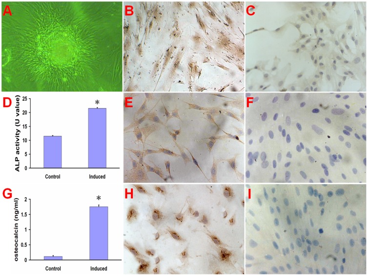
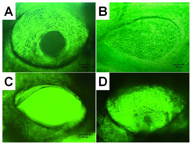
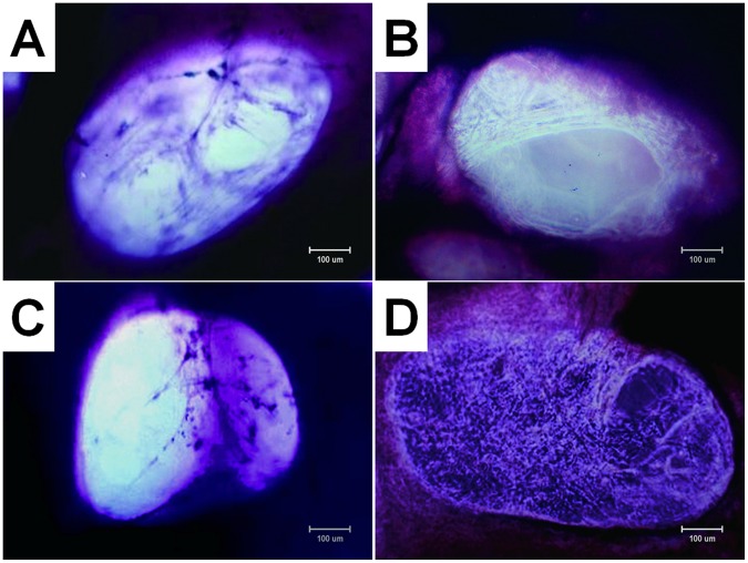
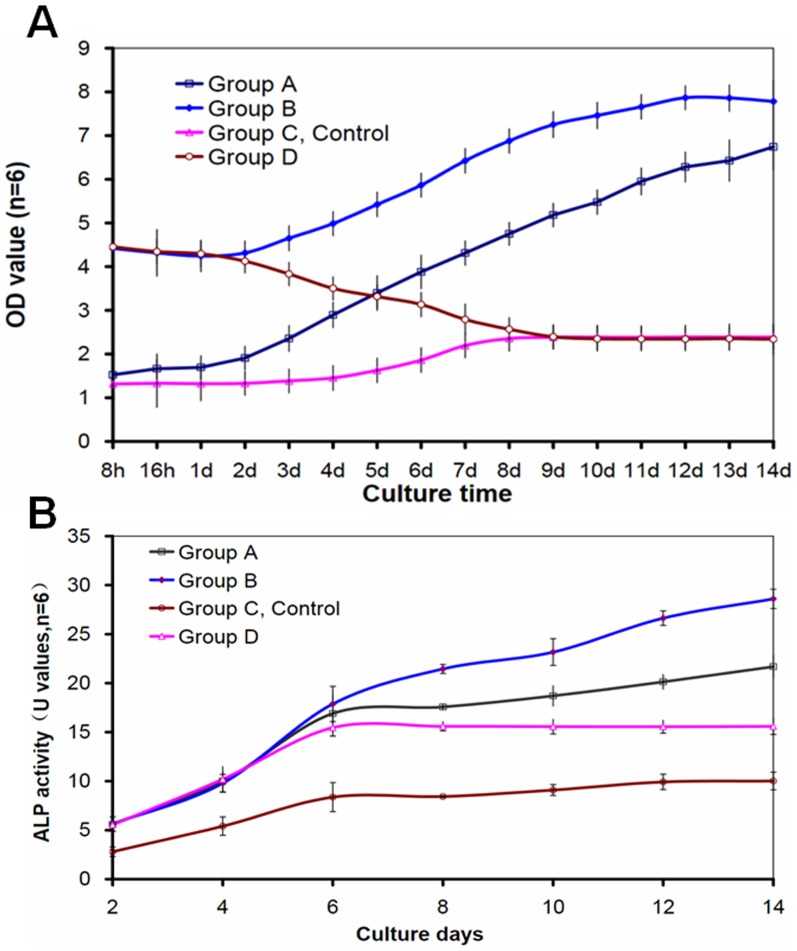
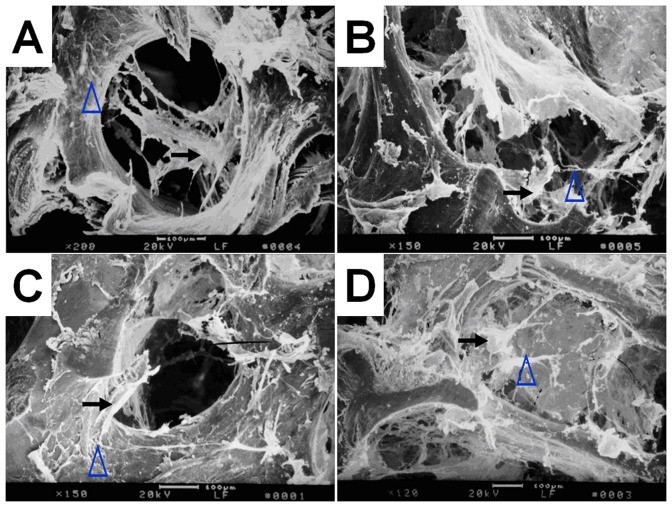
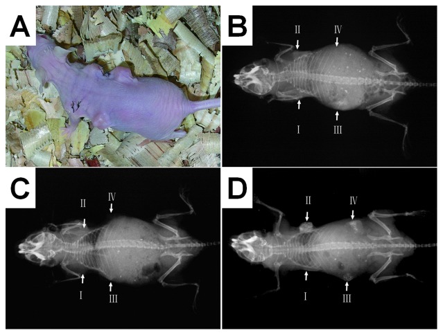
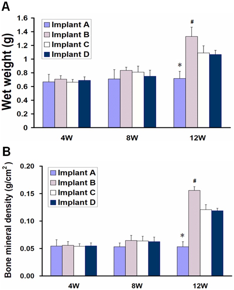
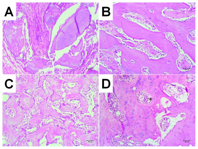
This study aimed to study the effects of initial cell density and in vitro culture method on the construction of tissue-engineered bone grafts and osteogenic activities. Human mesenchymal stem cells (hMSCs) were seeded onto cubic scaffolds prepared from demineralized bone matrix (DBM) by three methods - static, hydrodynamic, or fibrin hydrogel-assisted seeding. The resulting cell-scaffold constructs were cultured in vitro by static flask culture or hydrodynamic culture. The initial cell density and the subsequent in vitro proliferation and alkaline phosphate activities of the constructs were analyzed. The constructs were also subcutaneously implanted in nude mice to examine their in vivo osteogenic activities. Hydrogel-assisted seeding gave the highest seeding efficiency, followed by hydrodynamic and conventional static seeding. During in vitro culture, hydrodynamic culture produced higher plateau cell densities, alkaline phosphatase (ALP) activities, and extracellular matrix production than static culture. After subcutaneous implantation in nude mice, the implants prepared by the combination of hydrogel-assisted seeding and hydrodynamic culture produced higher wet weight and bone mineral density than implants prepared by other methods. The results suggest that the hydrogel-assisted seeding can substantially increase the initial seed cell density in scaffolds. Subsequent hydrodynamic culture can promote the proliferation and osteoblastic differentiation of the seeded cells. Correspondingly, bone grafts produced by the combination of these two methods achieved the highest osteogenic activity among the three methods employed.


Conflict of interest statement

Competing Interests: The authors have declared that no competing interests exist.

Figures

Figure 1
Figure 1. The culture and characterization of hMSCs.
The hMSCs formed calcium nodus after 12 day culture (A). The hMSCs (200×) stained immunohistochemically positive for ALP (B), osteocalcin (E) and collagen type I (H) compared to non-induced cells (C, F, and I). The ALP activity (D) of hMSCs and osteocalcin concentration (G) in the culture medium were significant higher in induced group than that in control group (non-induced cells). *p < 0. 01, compared with control group.
Figure 2
Figure 2. Phase-contrast photomicrographs (×100) of cell-scaffold constructs after in vitro culture for 12 d; (A) group A (dynamic seeding and dynamic culture), (B) group B (hydrogel-assisted seeding and static flask culture, (C) group C (static seeding and static flask culture, control group), and (D) group D (hydrogel-assisted seeding and dynamic culture).
Bar lengths are 100 um.
Figure 3
Figure 3. Photomicrographs (×100, methyl violet staining) of cell-scaffold constructs after in vitro culture for 12 d.
The number of attached cells and density of extracellular matrix (ECM) fibers in the interior of the scaffold are obvious different among four groups, with group B (B) > group D (D) > group A (A) > group C (C). Bar lengths are 100 um.
Figure 4
Figure 4. Proliferation of seeded cells in cell-scaffold constructs was detected by cell counting kit-8 (A) and osteoblastic differentiation of seeded cells in cell-scaffold constructs was evaluated by ALP activities (B).
The number of cells was increased with culture time except group C. The dynamic culture (groups A and B) showed an obvious ability of promoting proliferation of cells. The ALP activities in all groups increased from day 2 to day 14 (B). The ALP activities in groups A, B, D were statistically higher than that in groups C(p<0.05) from day 4 to day 14. indicates a statistically higher value compared with group C(p<0.05).
Figure 5
Figure 5. Scanning electron micrographs of cell-scaffold constructs after in vitro culture for 12 days.
The attached cells and extracellular matrix (ECM) fibers presented on the scaffolds in group B (B) and group D (D) are significantly outnumber those in group A (A) as well as group C (C).Bar lengths are 100 um. The black arrows indicate cells and the blue arrows indicate ECM fibers.
Figure 6
Figure 6. Nude mice subcutaneous implantation model for the evaluation of osteogenic activity; (A) a photograph showing a nude mouse with four implants; (B) a radiograph 4 weeks after implantation; (C) a radiograph 8 weeks after implantation; (D) a radiograph 12 weeks after implantation.
The radiographic densities of the implants increased from week 4 to week 12. The osteogenesis of implants was not clear at weeks 4 and 8 postoperative. It was not until 12 weeks postoperative that the imagings of implants in the radiographs were clearly observed. At week 12, implant II clearly showed increased density indicating calcification.
Figure 7
Figure 7. Wet weight and bone mineral density of implants after subcutaneous implantation in nude mice.
At 12 weeks postoperative, implant in group II showed higher wet weight (A) and bone mineral density (B) than that in other groups(p<0.05). *indicates a statistically significantly lower value compared with other implants; # indicates a statistically higher value compared with other implants.
Figure 8
Figure 8. HE staining of ectopic bone formation in nude mice at 12 weeks (×100), Implant I can be seen partially degraded DBM stand, surrounded by fibrous connective tissue replaced; Implant II showed more mature bone structure of a small beam than other groups; both Implant III and IV showed small beam structure of bone with some cartilage-like structure partially visible, bone formation maturity lower than Implant II.

Similar articles

Cited by

References

    1. Aubin H, Nichol JW, Hutson CB, Bae H, Sieminski AL, et al. (2010) Directed 3D cell alignment and elongation in microengineered hydrogels. Biomaterials 31: 6941–6951. - PMC - PubMed
    1. Chan BP, Leong KW (2008) Scaffolding in tissue engineering: general approaches and tissue-specific considerations. Eur Spine J 17 Suppl 4: 467–479. - PMC - PubMed
    1. Ouyang A, Yang ST (2007) Effects of mixing intensity on cell seeding and proliferation in three-dimensional fibrous matrices. Biotechnol Bioeng 96: 371–380. - PubMed
    1. Zhang ZY, Teoh SH, Chong WS, Foo TT, Chng YC, et al. (2009) A biaxial rotating bioreactor for the culture of fetal mesenchymal stem cells for bone tissue engineering. Biomaterials 30: 2694–2704. - PubMed
    1. Seebach C, Schultheiss J, Wilhelm K, Frank J, Henrich D (2010) Comparison of six bone-graft substitutes regarding to cell seeding efficiency, metabolism and growth behaviour of human mesenchymal stem cells (MSC) in vitro. Injury 41: 731–738. - PubMed

Publication types