. 2013;8(1):e53701.
doi: 10.1371/journal.pone.0053701. Epub 2013 Jan 11.

Sox2 is an androgen receptor-repressed gene that promotes castration-resistant prostate cancer

Affiliations

Sox2 is an androgen receptor-repressed gene that promotes castration-resistant prostate cancer

Steven Kregel et al. PLoS One. 2013.

Abstract

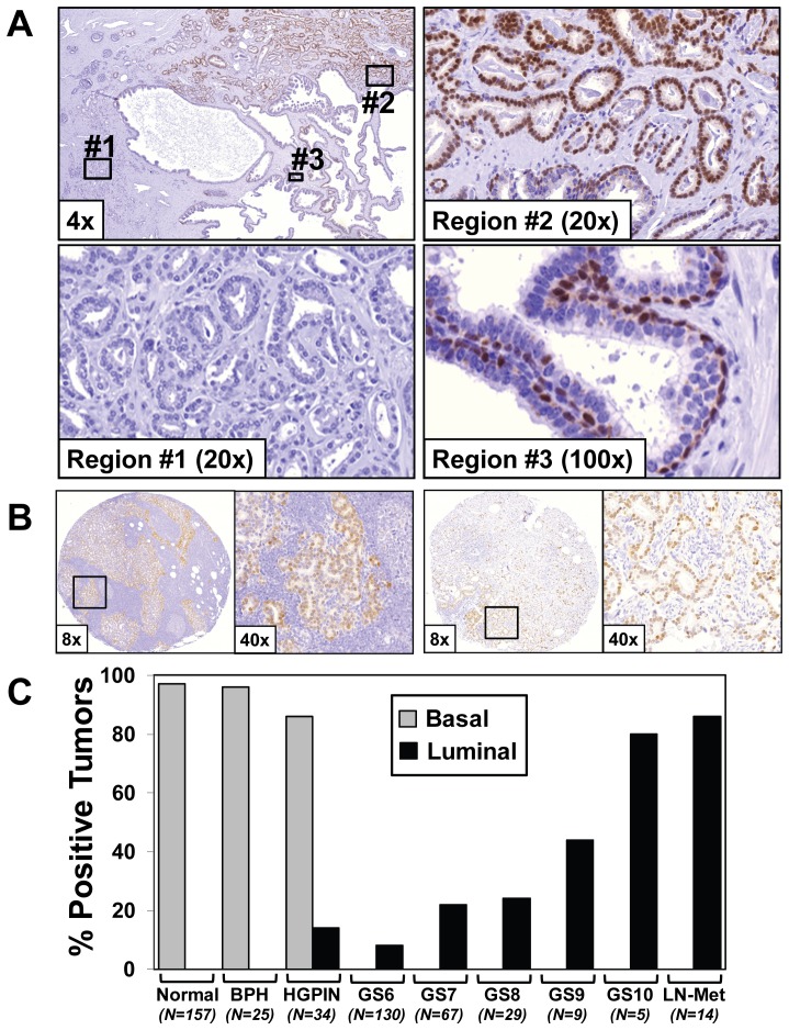
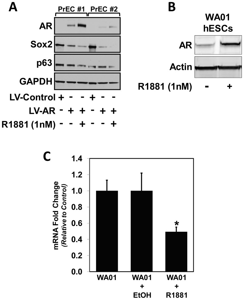
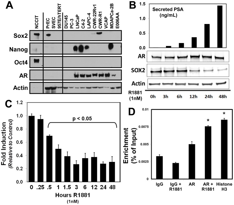
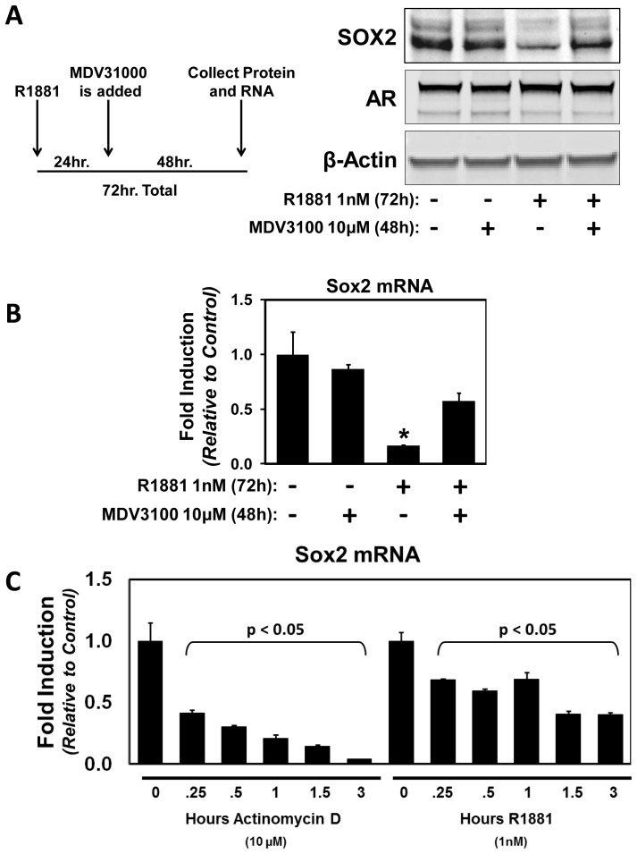
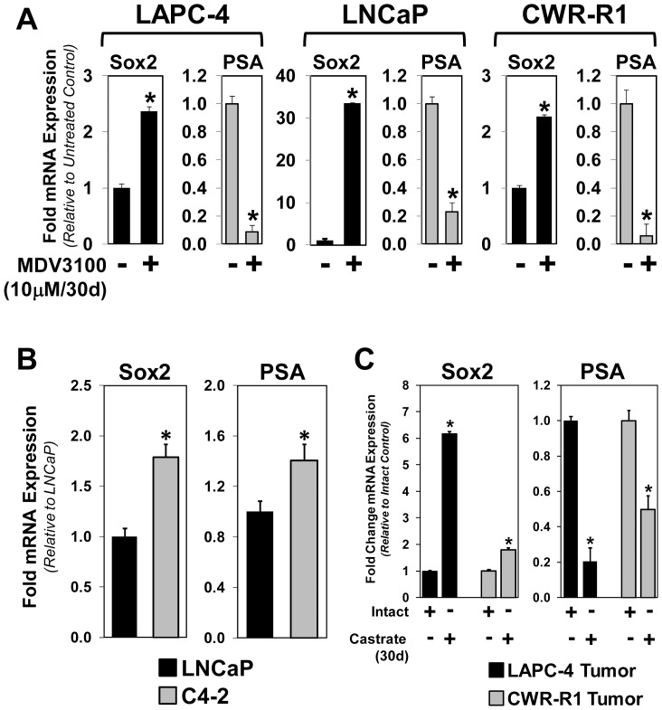
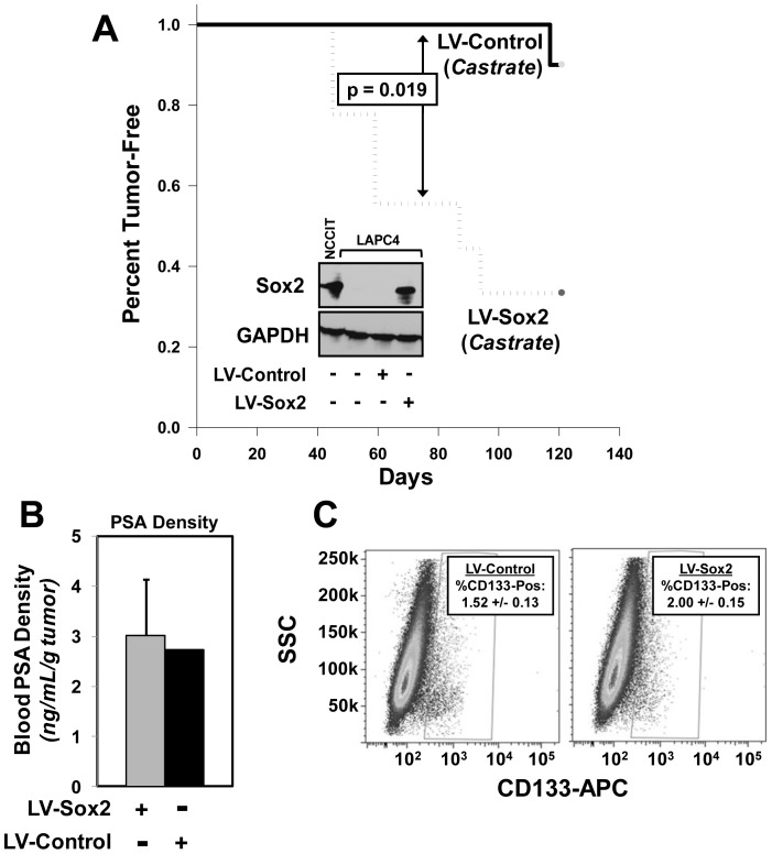
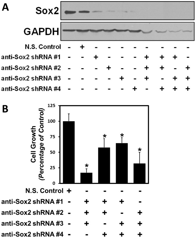
Despite advances in detection and therapy, castration-resistant prostate cancer continues to be a major clinical problem. The aberrant activity of stem cell pathways, and their regulation by the Androgen Receptor (AR), has the potential to provide insight into novel mechanisms and pathways to prevent and treat advanced, castrate-resistant prostate cancers. To this end, we investigated the role of the embryonic stem cell regulator Sox2 [SRY (sex determining region Y)-box 2] in normal and malignant prostate epithelial cells. In the normal prostate, Sox2 is expressed in a portion of basal epithelial cells. Prostate tumors were either Sox2-positive or Sox2-negative, with the percentage of Sox2-positive tumors increasing with Gleason Score and metastases. In the castration-resistant prostate cancer cell line CWR-R1, endogenous expression of Sox2 was repressed by AR signaling, and AR chromatin-IP shows that AR binds the enhancer element within the Sox2 promoter. Likewise, in normal prostate epithelial cells and human embryonic stem cells, increased AR signaling also decreases Sox2 expression. Resistance to the anti-androgen MDV3100 results in a marked increase in Sox2 expression within three prostate cancer cell lines, and in the castration-sensitive LAPC-4 prostate cancer cell line ectopic expression of Sox2 was sufficient to promote castration-resistant tumor formation. Loss of Sox2 expression in the castration-resistant CWR-R1 prostate cancer cell line inhibited cell growth. Up-regulation of Sox2 was not associated with increased CD133 expression but was associated with increased FGF5 (Fibroblast Growth Factor 5) expression. These data propose a model of elevated Sox2 expression due to loss of AR-mediated repression during castration, and consequent castration-resistance via mechanisms not involving induction of canonical embryonic stem cell pathways.


Conflict of interest statement

Competing Interests: The authors have declared that no competing interests exist.

Figures

Figure 1
Figure 1. Sox2 is uniformly expressed in a subset of hormone naïve prostate tumors and castration-resistant metastases.
A) Immunohistochemical staining of Sox2 demonstrating representative nuclear basal epithelial staining (dark red) in normal glands (Region #3), and two distinct tumor regions that are either uniformly Sox2-positve (Region #2) or Sox2-negative (Region #1). B) Expression of Sox2 in representative castration-resistant metastatic lesions. The box in the 8× magnification corresponds to the region shown in the 40× image. C) Percentage and distribution of Sox2 expression among prostate disease states. In normal, BPH, and HGPIN tissues, Sox2 is expressed in basal-epithelial cells (Grey Bars). Positive expression is defined by more than 50% of the cancer cells positive with a relative intensity of 1 or more on a scale of 0–3. In cancer tissues and metastases, Sox2 is either uniformly expressed or absent, and the percentage of Sox2-positive tumors increases with Gleason Score (Black Bars). BPH: Benign Prostatic Hyperplasia; HGPIN: high-grade prostatic intraepithelial neoplasia; GS: Gleason Score (N = number of individual patient specimens analyzed).
Figure 2
Figure 2. Sox2 is expressed in the majority of normal basal-epithelial cells, and cultures derived from prostate epithelial cells (PrECs) are either uniformly Sox2-Positive or Negative.
A) Immunofluorescent co-staining of Sox2 (green) with the basal-specific marker p63 (red), showing that 75% of normal basal-epithelial cells are positive for both Sox2 and p63 (yellow) while the remaining 25% are Sox2-negative (red). DAPI staining highlights nuclei (representative images of normal prostate epithelium acquired from three different individual patient specimens). B) Western blotting of a series of patient-derived Prostate (PrEC) and Seminal Vesicle (SVEC) epithelial sell (PrEC) cultures demonstrates that a portion of these cultures express detectable Sox2, while other PrEC and all SVEC cultures do not. C) Immunocytochemical staining of Sox2 showing uniform nuclear Sox2 expression in Sox2-positive PrECs and lack of Sox2-positive cells within Sox2-negative PrECs (representative images of three independent experiments).
Figure 3
Figure 3. Sox2 is negatively regulated by Androgen Receptor (AR) signaling in prostate epithelial cells (PrECs) and human embryonic stem cells (hESCs).
A) To test whether AR signaling impacted Sox2 expression, we ectopically expressed AR (LV-AR) and control lentivirus (LV-Control) in two different Sox2-positive PrEC lines (#1 and #2). Western blotting shows that expression and further ligand activation of AR using 1 nM R1881 results in decreased Sox2 protein expression and decreased p63 expression. GAPDH was used as a loading control. B) To test whether AR-mediated inhibition of Sox2 expression was specific to PrECs or could also occur in Sox2-positive human ES cells, we treated the WA01(H1) human ES cell line with androgen (1 nM R1881). Western blotting documents detectable endogenous AR expression in hESCs, which increases in response to androgen. β-Actin was used as a loading control. C) AR activation in hESCs results in a significant decrease in Sox2 mRNA expression as measured by qPCR (* indicates p<0.05). This was compared to untreated and vehicle treated cells (EtOH).
Figure 4
Figure 4. Androgen Receptor (AR) directly represses Sox2 expression in castration-resistant CWR-R1 cells.
A) Western blot of a panel of non-malignant and prostate cancer cell lines for Sox2, Nanog, Oct4, and AR. β-Actin was used as a loading control. Expression of Sox2 in castration-resistant CWR cells is not accompanied by co-expression of Nanog or Oct4. LNCaP, C4-2B, LAPC-4, and MDA-PCa2B cells expressed detectable Nanog, which is presumably the NanogP8 retrogene (Jeter et al., 2011). The human embryonal carcinoma cell line NCCIT was used as a positive control for Sox2, Nanog, and Oct4 at a 1∶10 dilution. B) Decreased expression of Sox2 upon AR stimulation with physiologic levels of androgen (1 nM R1881) in castration-resistant CWR-R1 prostate cancer cells. Protein lysates from cells treated at defined intervals (3–48 hours) after androgen treatment were subjected to western blotting, and accumulation of secreted PSA expressed in the media validates increased AR signaling. C) Rapid decrease of Sox2 mRNA in CWR-R1 cells upon AR stimulation as measured by qPCR. Levels at 0.5 hrs and beyond represent a statistically significant decrease in Sox2 mRNA (p<0.05). D) AR Chromatin Immunoprecipitation (ChIP) documents direct binding of ligand-activated AR to the Sox2 enhancer region in response to AR stimulation by R1881. CWR-R1 cells were treated with vehicle control or 1 nM R1881, and enrichment of the Sox2 promoter after AR-ChIP was normalized as a percentage of total chromatin input. IgG and Histone H3 served as negative and positive controls, respectively. When compared to total input, both the positive control Histone H3 and ligand-activated AR significantly enriched for the Sox2 enhancer (p<0.05). Data represents three independent experiments.
Figure 5
Figure 5. Androgen Receptor-mediated repression of Sox2 expression can be reversed by treatment with the Anti-Androgen MDV3100.
A) To verify that the decrease of Sox2 protein is specific to AR activation and can be reversed by an AR antagonist, CWR-R1 cells were grown in 1 nm R1881 or vehicle for 24 hrs, and then either 10 µM MDV3100 or vehicle was added to the culture medium for an additional 48 hrs. Western blots show a decrease in Sox2 protein with R1881 that can be returned to basal levels with addition of MDV3100, without any change in AR protein. β-Actin was used as a loading control. A schematic outlines the time frame of these experiments. B) A decrease of Sox2 mRNA with R1881 treatment was measured using qPCR (*p<0.05), which was brought back to control levels upon treatment with MDV3100. C) A time course of treatment with a known inhibitor of transcription, 10 µM Actinomycin D, yielded a similar rapid decrease of Sox2 mRNA in CWR-R1 cells upon AR stimulation with R1881 as measured by qPCR. Levels at 0.25 hrs and beyond represent a statistically significant decrease in Sox2 mRNA (p<0.05), showing similar kinetics of transcriptional repression with both R1881 and Actinomycin D drug treatments.
Figure 6
Figure 6. Sox2 expression is increased in MDV3100-resistant prostate cancer cells and castration-resistant prostate tumors.
A) To test whether Sox2 expression is associated with resistance to AR pathway inhibition, we developed a series of prostate cancer cell lines that were resistant to MDV3100. After 30 days of continuous treatment, MDV3100-resistant lines expressed significantly higher Sox2 as measured using qPCR (*p<0.05). AR-mediated PSA expression was also significantly reduced in these lines. B) Comparison between the castration-sensitive LNCaP and isogenic castration-resistant C4-2 cell lines documents a significant increase in Sox2 expression in castration-resistant C4-2 cells (*p<0.05). C) Increased Sox2 expression in castration-resistant xenografts of LAPC-4 and CWR-R1 tumors. Tumors were allowed to establish and then host mice were castrated; 30 days later castration-resistant tumors were harvested and total mRNA analyzed for Sox2 and PSA. These data show increased Sox2 within castration-resistant LAPC-4 and CWR-R1 tumors (*p<0.05; data represents quantitation from multiple tumor specimens).
Figure 7
Figure 7. Sox2 expression promotes castration resistant prostate cancer tumor formation.
A) Ectopic expression of Sox2 in castrate-sensitive LAPC-4 (LV-Sox2, n = 10 mice) cells is sufficient to promote tumor take in castrated male nude hosts compared to control mice (LV-Control, n = 10) (p = 0.019). Inset: western blot documenting ectopic lentiviral Sox2 expression in LAPC-4 cells. GAPDH was used as a loading control. NCCIT cells are used as a Sox2-positive control. B) Castrated LAPC-4-Sox2 versus LAPC-4-Control tumors do not have differences in serum PSA density. These data support that Sox2 expression does not confer a less-differentiated tumor phenotype. PSA density is the ng/mL total PSA per gram tumor. C) Expression of Sox2 in LAPC-4 does not significantly increase the percentage of rare CD133-positive cells, which are thought to be putative cancer stem/initiating cells.
Figure 8
Figure 8. Sox2 expression is necessary for the growth of castration-resistant prostate cancer cells.
A) Depletion of Sox2 protein expression using four different lentiviral shRNA constructs. To test the impact of inhibiting Sox2 expression in castration-resistant CWR-R1 cells, we used a series of shRNAs that targeted different regions of the Sox2 mRNA transcript. These data show that each shRNA sequence results in decreased Sox2 protein expression after 72 hours, and combinations of three were used to obtain levels below detection while controlling for potential off-target (i.e. non-Sox2 specific) effects. A non-silencing shRNA (NSC) control was used in comparison. GAPDH was used as a loading control. B) Decreased expression of Sox2 results in significant growth inhibition. Cell growth was measured using MTT reduction after five days in complete media. In all four instances of Sox2 knockdown, cell growth was significantly diminished compared to the non-silencing control.
Figure 9
Figure 9. Sox2 expression is associated with the expression of FGF5 and not established human embryonic stem cell Sox2 target genes.
A) The expression of 83 embryonic stem cell-associated genes was analyzed via quantitative real-time PCR analyses to identify Sox2-associated gene changes in LAPC-4-Sox2 cells (compared to LAPC-4-Control cells). Open bars represent genes that are previously identified Sox2-target genes in hESCs (Boyer et al. 2005). No changes in any of these 18 genes were associated with Sox2 expression in LAPC-4 cells. Rather, significant changes in expression were detected for FGF5, Kit, NR5A2, PDX1, and RUNX2 (black bars). B) Elevated expression of FGF5 in castration-resistant LAPC-4 and CWR-R1 tumors (*p<0.05; same mRNA as described in Figure 6C).

Similar articles

Cited by

References

    1. Huggins C, Stevens RE, Hodges CV (1941) Studies on Prostate Cancer: II. The Effects of Castration on Advanced Carcinoma of the Prostate Gland. Archives of Surgery 43: 209–223.
    1. Isaacs JT, Isaacs WB (2004) Androgen receptor outwits prostate cancer drugs. Nat Med 10: 26–27. - PubMed
    1. Singh P, Uzgare A, Litvinov I, Denmeade SR, Isaacs JT (2006) Combinatorial androgen receptor targeted therapy for prostate cancer. Endocr Relat Cancer 13: 653–666. - PubMed
    1. Takahashi K, Yamanaka S (2006) Induction of pluripotent stem cells from mouse embryonic and adult fibroblast cultures by defined factors. Cell 126: 663–676. - PubMed
    1. Rodriguez-Pinilla SM, Sarrio D, Moreno-Bueno G, Rodriguez-Gil Y, Martinez MA, et al. (2007) Sox2: a possible driver of the basal-like phenotype in sporadic breast cancer. Mod Pathol 20: 474–481. - PubMed

Publication types

MeSH terms