. 2013;8(1):e53739.
doi: 10.1371/journal.pone.0053739. Epub 2013 Jan 11.

Desmoglein 2 is less important than desmoglein 3 for keratinocyte cohesion

Affiliations

Desmoglein 2 is less important than desmoglein 3 for keratinocyte cohesion

Eva Hartlieb et al. PLoS One. 2013.

Abstract

Desmosomes provide intercellular adhesive strength required for integrity of epithelial and some non-epithelial tissues. Within the epidermis, the cadherin-type adhesion molecules desmoglein (Dsg) 1-4 and desmocollin (Dsc) 1-3 build the adhesive core of desmosomes. In keratinocytes, several isoforms of these proteins are co-expressed. However, the contribution of specific isoforms to overall cell cohesion is unclear. Therefore, in this study we investigated the roles of Dsg2 and Dsg3, the latter of which is known to be essential for keratinocyte adhesion based on its autoantibody-induced loss of function in the autoimmune blistering skin disease pemphigus vulgaris (PV). The pathogenic PV antibody AK23, targeting the Dsg3 adhesive domain, led to profound loss of cell cohesion in human keratinocytes as revealed by the dispase-based dissociation assays. In contrast, an antibody against Dsg2 had no effect on cell cohesion although the Dsg2 antibody was demonstrated to interfere with Dsg2 transinteraction by single molecule atomic force microscopy and was effective to reduce cell cohesion in intestinal epithelial Caco-2 cells which express Dsg2 as the only Dsg isoform. To substantiate these findings, siRNA-mediated silencing of Dsg2 or Dsg3 was performed in keratinocytes. In contrast to Dsg3-depleted cells, Dsg2 knockdown reduced cell cohesion only under conditions of increased shear. These experiments indicate that specific desmosomal cadherins contribute differently to keratinocyte cohesion and that Dsg2 compared to Dsg3 is less important in this context.


Conflict of interest statement

Competing Interests: The authors have declared that no competing interests exist.

Figures

Figure 1
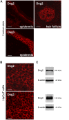
Figure 1. Expression patterns of Dsg2 and Dsg3 in human keratinocytes.
(A) Immunofluorescence staining of cryosections of human skin demonstrating expression of Desmoglein (Dsg) 2 and Dsg3 in epidermis. For Dsg2 also the expression in the hair follicle is shown. Scale bar, 20 µm. (B) Immunofluorescence staining of Dsg2 and Dsg3 in a human keratinocyte cell line (HaCaT). Cells were cultured for 4 d in high Ca2+-medium. Scale bar, 20 µm. (C) Western blots analysis of Dsg2 and Dsg3 in confluent 4 d HaCaT cells. ß-actin was used as loading control.
Figure 2
Figure 2. Antibody-mediated targeting of Dsg3, but not of Dsg2, led to profound loss of cell cohesion.
(A) After antibody incubation for 24 h with either a monoclonal Dsg2 antibody (Dsg2 mAb) or AK23, confluent HaCaT monolayers were subjected to dispase-based dissociation assays. Loss of cell cohesion was detectable in cells incubated with AK23 only. (n = 8; * p<0.05 vs. control) Photos were taken immediately after assay performance. 1 hour incubation with 5 mM EGTA led to profound loss of cell cohesion indicating the non-hyperadhesive state of HaCaT cells used for these experiments. (n = 6) (B) Exposing the cells to more severe mechanical stress in dissociation assays increased fragment numbers after AK23 treatment only. (n = 6; * p<0.05 vs. control) (C) Atomic force microscopy was used to demonstrate antibody-mediated interference with homophilic Dsg2 and Dsg3 binding. Both antibodies reduced the binding frequency of their respective antigens. (>1000 force distance cycles on more than 2 different cantilever/substrate combinations; * p<0.05 vs. control) (D) Dispase-based dissociation assay performed with confluent Caco-2 cells after 24 h incubation with Dsg2 mAb showed a significant increase in fragment numbers compared to control cells. (n≥20; * p<0.05 vs. control) (E) Protein expression of Dsg2 in Caco-2 cells was proven by immunofluorescence (scale bar, 20 µm) and (F) Western blot analysis. ß-actin was used as loading control.
Figure 3
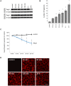
Figure 3. Depletion of Dsg3 is an event subsequent to loss of cell-cell adhesion after AK23 incubation.
(A) 24 hours after incubation with AK23 depletion of Dsg3 was detectable in the Triton-soluble but not in the Triton-insoluble fraction of HaCaT cell lysates. ß-actin was used as loading control. (n = 3) (B) Loss of cell cohesion started after 30 min of AK23 incubation as detected in the dispase-based dissociation assays. (n = 6; * p<0.05 vs. control) (C) Similarly, laser tweezers experiments to evaluate binding of Dsg3-coated microbeads on HaCaT cell surface showed a reduction of tightly attached beads starting after 30 min following AK23 exposure. (n = 6; * p<0.05 vs. control) (D) AK23 binding to the cell surface at all time points of laser tweezers measurements was demonstrated by immunostaining for mouse Fc after fixation. Scale bar, 20 µm.
Figure 4
Figure 4. SiRNA-mediated depletion of Dsg2 and Dsg3 in HaCaT cells.
(A) Efficient Dsg2 (red) depletion was detected by immunofluorescence staining in HaCaT cells. Actin filaments are colored in green with Alexa Fluor®488 phalloidin. Scale bar, 20 µm. (B) Immunofluorescence staining for Dsg3 (red) and F-actin (green) after siRNA-mediated Dsg3 silencing. Scale bar, 20 µm. (C) Immunoblot analysis demonstrated a decrease in protein expression of either Dsg2 or Dsg3 after respective siRNA-mediated knockdown whereas protein levels of Dsc2 and E-cadherin were not changed after siRNA-mediated silencing of Dsg2 or Dsg3. Dsg2 depletion induced a slight but significant reduction of Dsg3 protein content. In contrast, Dsg2 levels were unchanged after Dsg3 silencing. ß-actin was used as loading control and band density was normalized to ß-actin. (n = 6; * p<0.05 vs. n. t. siRNA) (D) In contrast to Dsg3, endogenous Dsg2 expression was primarily detectable in the desmosomal pool (left panel). SiRNA-mediated gene silencing caused a reduction of Dsg3 in both protein fractions. Desmoplakin was used to identify the Triton X-100-insoluble fraction as the desmosome containing pool. Under control conditions, band density analysis revealed a five times higher ratio of insoluble over soluble protein levels of Dsg2 compared to Dsg3 indicating a more pronounced extradesmosomal localization of Dsg3 (right panel). (n = 5).
Figure 5
Figure 5. Dsg3 but not Dsg2 depletion leads to loss of cell-cell adhesion.
(A) Culture wells photographed after performance of the dispase-based dissociation assay under indicated conditions. Loss of cell-cell adhesion was detectable after AK23 incubation and siRNA-induced Dsg3 knockdown, but absent when Dsg2 levels were reduced by siRNA. (B) Mean fragment numbers per well under experimental conditions when control monolayers withstood the mechanical stress level and stayed intact. (n>20; * p<0.05 vs. n. t. siRNA) (C) Fragment numbers after exposing cell monolayers to higher mechanical stress. (n≥5; * p<0.05 vs. n. t. siRNA).

Similar articles

Cited by

References

    1. Waschke J (2008) The desmosome and pemphigus. Histochem Cell Biol 130: 21–54. - PMC - PubMed
    1. Getsios S, Huen AC, Green KJ (2004) Working out the strength and flexibility of desmosomes. Nat Rev Mol Cell Biol 5: 271–281. - PubMed
    1. Tsang SM, Brown L, Lin K, Liu L, Piper K, et al. (2012) Non-junctional human desmoglein 3 acts as an upstream regulator of Src in E-cadherin adhesion, a pathway possibly involved in the pathogenesis of pemphigus vulgaris. J Pathol 227: 81–93. - PubMed
    1. Nie Z, Merritt A, Rouhi-Parkouhi M, Tabernero L, Garrod D (2010) Membrane-impermeable Cross-linking Provides Evidence for Homophilic, Isoform-specific Binding of Desmosomal Cadherins in Epithelial Cells. Journal of Biological Chemistry 286: 2143–2154. - PMC - PubMed
    1. Stanley JR, Amagai M (2006) Pemphigus, bullous impetigo, and the staphylococcal scalded-skin syndrome. N Engl J Med 355: 1800–1810. - PubMed

Publication types

LinkOut - more resources