. 2013;8(1):e53884.
doi: 10.1371/journal.pone.0053884. Epub 2013 Jan 9.

Asymptomatic cattle naturally infected with Mycobacterium bovis present exacerbated tissue pathology and bacterial dissemination

Affiliations

Asymptomatic cattle naturally infected with Mycobacterium bovis present exacerbated tissue pathology and bacterial dissemination

Álvaro Menin et al. PLoS One. 2013.

Abstract

Rational discovery of novel immunodiagnostic and vaccine candidate antigens to control bovine tuberculosis (bTB) requires knowledge of disease immunopathogenesis. However, there remains a paucity of information on the Mycobacterium bovis-host immune interactions during the natural infection. Analysis of 247 naturally PPD+ M. bovis-infected cattle revealed that 92% (n = 228) of these animals were found to display no clinical signs, but presented severe as well as disseminated bTB-lesions at post-mortem examination. Moreover, dissemination of bTB-lesions positively correlated with both pathology severity score (Spearman r = 0.48; p<0.0001) and viable tissue bacterial loads (Spearman r = 0.58; p = 0.0001). Additionally, granuloma encapsulation negatively correlated with M. bovis growth as well as pathology severity, suggesting that encapsulation is an effective mechanism to control bacterial proliferation during natural infection. Moreover, multinucleated giant cell numbers were found to negatively correlate with bacterial counts (Spearman r = 0.25; p = 0.03) in lung granulomas. In contrast, neutrophil numbers in the granuloma were associated with increased M. bovis proliferation (Spearman r = 0.27; p = 0.021). Together, our findings suggest that encapsulation and multinucleated giant cells control M. bovis viability, whereas neutrophils may serve as a cellular biomarker of bacterial proliferation during natural infection. These data integrate host granuloma responses with mycobacterial dissemination and could provide useful immunopathological-based biomarkers of disease severity in natural infection with M. bovis, an important cattle pathogen.


Conflict of interest statement

Competing Interests: The authors have declared that no competing interests exist.

Figures

Figure 1
Figure 1. Bacteriology analysis and molecular typing of M. bovis.
(A) Tissue homogenate obtained from PPD+ asymptomatic animals was inoculated in Ogawa-Kudoh+sodium pyruvate and incubated at 37°C for 8 weeks. After that, colonies were counted and DNA extraction method employed. Purified DNA obtained from (A) was used as template for PCR amplification of (B) IS1081 (∼135 bp) or (C) RvD1Rv2031c (∼500 bp) gene sequences. Amplification products of single PCR from representative samples are shown. Lane M: 100 bp DNA ladder; lanes 1–22 PCR products of M. bovis isolates; AN5 - Mycobacterium bovis AN5 strain, standard strain positive control; 37Rv – Mycobacterium tuberculosis strain, H37Rv; D4 - Mycobacterium avium Subsp. avium D4 strain, non-tuberculosis mycobacteria (NMTBC) member; lane NC, negative control (without DNA).
Figure 2
Figure 2. Clinical and gross pathology findings in cattle naturally infected with M. bovis.
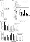
(A) Following clinical examination, animals bTB-positive (n = 247) were categorized according to their clinical status into asymptomatic (AS, n = 228), moderate symptoms (MS, n = 11), severe symptoms (SS, n = 8). (B) Gross pathology analysis further divided the PPD+ asymptomatic animals into two groups: the presence of visible bTB-lesions (VL) or absence of visible bTB-lesions (NVL). (C and D) Severity of tissue gross pathology in asymptomatic bTB bovines was scored by applying a previously described semi-quantitative scoring system (Vordermeier et al, 2002). Results shown are median of scores ± SEM. (C) Organs/Tissues: thoracic organs and tissues (lung (LG), pleura (PLE), pericardia (PC)); abdominal (liver (LV), spleen (SP), intestine (IN), mesentery (MT), genitor-urinary system (GS), as well as udder (UD), other tissues (OT)); (D) Lymph nodes: head lymph nodes (mandibular (ML), parotid (PL), retropharyngeal (RL) lymph nodes and palatine tonsil (PT)); thoracic lymph nodes (tracheobronchial (TL), bronchial (BL) and mediastinal (MDL)); abdominal lymph nodes (hepatic (HL), mesenteric (MSL)) as well as Iliac (ILL), Sciatic (SL), pre-scapular (PEL) and pre-crural (PCL) lymph nodes. (E) Frequency of bTB-lesions in different organs/lymph nodes affected of asymptomatic bTB bovines. Legends as described in (C) and (D). (E, inset) Frequency of lung lobes affected (left cranial lobe (LCR), left caudal lobe (LCA), right cranial lobe (RCR), right caudal lobe (RCA), middle lobe (MD) and accessory lobe (AC) were determined.
Figure 3
Figure 3. Anatomical dissemination of bTB-lesions and infection burden in asymptomatic cattle naturally infected with M. bovis.
(A) Frequency of animals categorized according to their anatomical dissemination of bTB-lesions into five levels: I, lesions of bTB in the head lymph nodes, including retropharyngeal, mandibular, and parotid lymph nodes; II, presence of lesions of bTB in thoracic lymph nodes, including the mediastinal, bronchial, tracheobronchial lymph nodes, or in abdominal lymph nodes, including mesenteric, inguinal, gastric, hepatic, splenic, renal, sub-iliac, medial and lateral iliac lymph nodes; III, simultaneous presence of lesions suggestive of bTB in thoracic and abdominal lymph nodes; IV, presence of lesions of bTB in organs of the thoracic or abdominal cavity; and V, simultaneous presence of lesions of bTB in organs of thoracic and abdominal cavities. Schematic cartoon of the scoring system is represented. Continuous line represents both cavities affected (thoracic and abdominal); dotted line represents only one cavity affected. Green balloons indicate affected organs and blue balloons indicate affected lymph nodes. Correlation between levels of lesion dissemination and pathology severity (B) or mycobacterial loads (C) in cattle naturally infected with M. bovis. In (B), the results are expressed applying the gross pathology severity semi-quantitative scoring per animal previously described in , and (C) as number of CFU.mL−1 (colony-forming units per mL of granulomatous tissue homogenate). Spearman’s correlation indexes (Spearman’s r and p values) are shown in the graphs.
Figure 4
Figure 4. Histopathological analysis of granuloma encapsulation in asymptomatic cattle naturally infected with
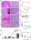
M. bovis. (A) Granulomas were formalin-fixed, paraffin-embedded and 4 µm-sections Massońs trichrome stained, categorized and scored according to their intensity of encapsulation of primary granulomas into three levels: (1 and 2) I, thin encapsulation; (3 and 4) II, dense fibrous capsule; and (5 and 6) III, thickly fibrous encapsulation. (A, inset) Acid Fast-Bacilli (AFB). (Left panels, slides shown at 10× magnification; Scale bar  = 100 µm. Right panels, slides shown at 40× magnification; Scale bar  = 50 µm. Inset, slides shown at 100× magnification. Correlation between intensity of granuloma encapsulation with mycobacterial loads (B) or gross pathology severity score (C) in cattle naturally infected with M. bovis are presented. Spearman’s correlation indexes (Spearman’s r and p values) are shown in the graphs.
Figure 5
Figure 5. Histopathological analysis of cellular profile of granulomatous response in lung of asymptomatic cattle naturally infected with M. bovis.
(A) Lung tissues were categorized according to the granuloma cellular response profile and tissue remodeling into four groups: I-IV. Representative lung-tuberculous granulomatous response patterns are shown: I (1 and 2) encapsulated granulomas with caseous necrosis areas and presence of several scattered lymphocytes and dense clusters of neutrophils near the capsule; II (3 and 4) encapsulated granuloma, with extensive areas of caseous necrosis. Granulomatous cellular response composed primarily of epithelioid macrophages, lymphocytes, multinucleated Langhańs giant cells and clusters of neutrophils; III (5 and 6) encapsulated granulomas, with extensive multicentric areas of caseous necrosis and centralized dystrophic mineralization. Granulomatous cellular response composed of epithelioid macrophages and scattered Langhan’s giant cells, which surround the necrotic areas with dense clusters of lymphocytes and few neutrophils; III (7 and 8) encapsulated granulomas, with extensive multicentric areas of caseous necrosis and centralized dystrophic mineralization. Granulomatous cellular response composed of epithelioid macrophages admixed with increased numbers of multinucleated Langhan’s giant cells, dense clusters lymphocytes and few neutrophils. Left panels, slides shown at 10× magnification; Scale bar  = 100 µm. Right panels, slides shown at 40× magnification; Scale bar  = 50 µm. (B) Results presented are mean ± SEM for each group shown in (A). Correlation between neutrophil counts and histopathology grades (C), Correlation between viable mycobacterial loads and neutrophils (D), multinucleated Langhańs giant cells counts (E) as well as lung-granulomatous response profile (F). Spearman’s correlation indexes (Spearman’s r and p values) are shown in the graphs (n = 168 animals for correlation figures).

Similar articles

Cited by

References

    1. World Health Organization (2008) Global tuberculosis control - surveillance, planning, financing: WHO report 2008. WHO/HTM/TB/2008 393: 1–304.
    1. Thoen C, LoBue P, de Kantor I (2006) The importance of Mycobacterium bovis as a zoonosis. Vet Microbiol 112: 339–3345. - PubMed
    1. Cosivi O, Grange JM, Daborn CJ, Raviglione MC, Fujikura T, et al. (1998) Zoonotic tuberculosis due to Mycobacterium bovis in developing countries. Emerg Infect Dis 4: 59–70. - PMC - PubMed
    1. de Kantor IN, Ambroggi M, Poggi S, Morcillo N, Telles MADS, et al. (2008) Human Mycobacterium bovis infection in ten Latin American countries. Tuberculosis 88: 358–365. - PubMed
    1. Berg S, Firdessa R, Habtamu M, Gadisa E, Mengistu A, et al. (2009) The Burden of Mycobacterial Disease in Ethiopian Cattle: Implications for Public Health. PLoS One 4: e5068 doi:10.1371/journal.pone.0005068 - DOI - PMC - PubMed

Publication types

LinkOut - more resources