. 2013;8(1):e53893.
doi: 10.1371/journal.pone.0053893. Epub 2013 Jan 10.

Scanning electron microscopy-based approach to understand the mechanism underlying the adhesion of dengue viruses on ceramic hydroxyapatite columns

Affiliations

Scanning electron microscopy-based approach to understand the mechanism underlying the adhesion of dengue viruses on ceramic hydroxyapatite columns

Maiko Saito et al. PLoS One. 2013.

Abstract

Although ceramic hydroxyapatite (HAp) chromatography has been used as an alternative method ultracentrifugation for the production of vaccines, the mechanism of virus separation is still obscure. In order to begin to understand the mechanisms of virus separation, HAp surfaces were observed by scanning electron microscopy after chromatography with dengue viruses. When these processes were performed without elution and with a 10-207 mM sodium phosphate buffer gradient elution, dengue viruses that were adsorbed to HAp were disproportionately located in the columns. However, when eluted with a 10-600 mM sodium phosphate buffer gradient, few viruses were observed on the HAp surface. After incubating the dengue viruses that were adsorbed on HAp beads at 37°C and 2°C, the sphericity of the dengue viruses were reduced with an increase in incubation temperature. These results suggested that dengue virus was adsorbed to the HAp surface by electronic interactions and could be eluted by high-salt concentration buffers, which are commonly used in protein purification. Furthermore, virus fusion was thought to occur with increasing temperature, which implied that virus-HAp adhesion was similar to virus-cell adhesion.


Conflict of interest statement

Competing Interests: Maiko Saito and Yae Kurosawa are employees by HOYA Corporation. This does not alter the authors’ adherence to all the PLOS ONE policies on sharing data and materials.

Figures

Figure 1
Figure 1. Illustration of experimental methods.
A: The flow direction and the division of the hydroxyapatite (HAp) column into 3 parts. After removing the HAp particles from the column, HAp particles were collected in each part of the column. Column head: lower position of the column (upstream side); Mid-column: middle position of the column; Column end: upper position of the column (downstream side). B: Virus elution and incubation processes of HAp columns. Detailed conditions are described in the Materials and methods, section 3–6.
Figure 2
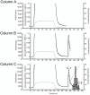
Figure 2. HAp chromatogram of dengue virus culture fluids from 3 different elution processes.
The solid line is the UV absorbance at 280 nm, the broken line is the UV absorbance at 260 nm, and the dashed line is the conductivity of the elution. The shaded histogram indicates the hemagglutination unit of each fraction. Column A: No elution; Column B: 10–207 mM NaPB (pH 7.2) gradient elution; Column C: 10–600 mM NaPB (pH 7.2) gradient elution. C-1: Admixture proteins peak; C-2: Virus peak.
Figure 3
Figure 3. SEM images of control HAp surfaces. A: Untreated HAp particles; B: HAp particles after loading of cell culture fluid (virus-free).
Figure 4
Figure 4. Observation of HAp surfaces that were used in the 3 different elution processes.
Column A: No elution; Column B: 10–207 mM NaPB (pH 7.2) gradient elution; Column C: 10–600 mM NaPB (pH 7.2) gradient elution. Each column was divided into 3 sections (Column end, Mid-column, Column head). The white triangles indicate dengue viruses.
Figure 5
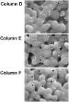
Figure 5. Observation of HAp surfaces that were incubated under 3 different conditions.
Column D: no incubation; Column E: incubated at 2°C for 7 days; Column F: incubated at 37°C for 7 days. HAp particles of the column head were observed with scanning electron microscopy. The white triangles indicate spherical-shaped viruses. The black triangles indicate flat-shaped viruses.

Similar articles

Cited by

References

    1. Lambert DM, Pons MW, Mbuy GN, Dorsch-Hasler K (1980) Nucleic acids of respiratory syncytial virus. J Virol 36: 837–846. - PMC - PubMed
    1. Morenweiser R (2005) Downstream processing of viral vectors and vaccines. Gene Ther Suppl 1: S103–10. - PubMed
    1. Song Z, Dong C, Wang L, Chen DE, Bi G, et al. (2010) A novel method for purifying bluetongue virus with high purity by co-immunoprecipitation with agarose protein A. Virol J. 7: 126. - PMC - PubMed
    1. Iyer G, Ramaswamy S, Asher D, Mehta U, Leahy A, et al. (2011) Reduced surface area chromatography for flow-through purification of viruses and virus like particles. J Chromatogr A 1218: 3973–3981. - PubMed
    1. Sugo K, Ogawa T (2007) Three-dimensional culture of rat bone marrow cells using hydroxyapatite microcarrier. Tiss Cult Res Commun 26: 125–131.

LinkOut - more resources