. 2013;8(1):e53918.
doi: 10.1371/journal.pone.0053918. Epub 2013 Jan 10.

Profilin 1 as a target for cathepsin X activity in tumor cells

Affiliations

Profilin 1 as a target for cathepsin X activity in tumor cells

Urša Pečar Fonović et al. PLoS One. 2013.

Abstract

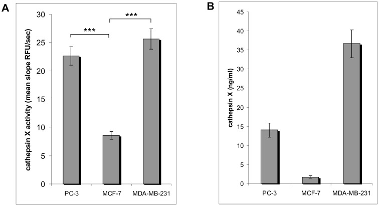
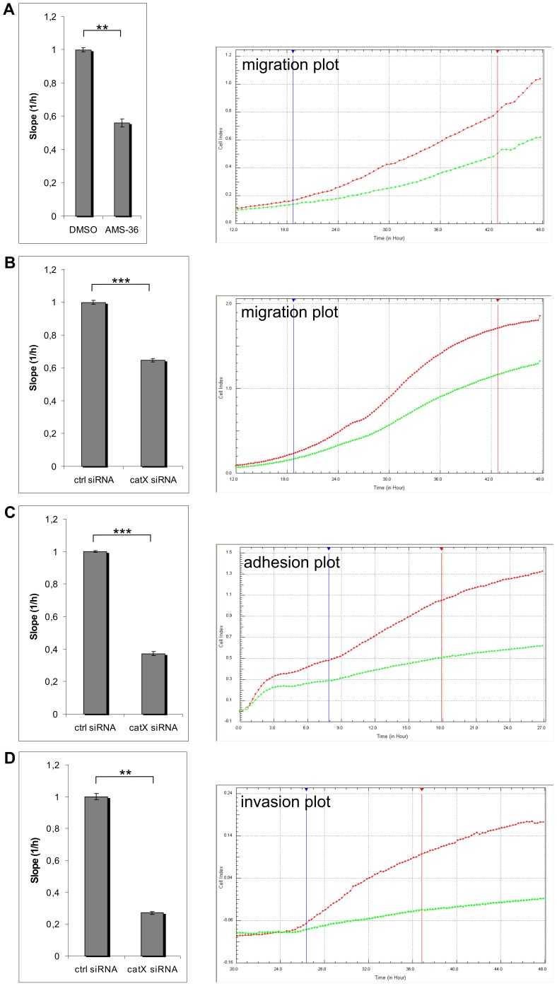
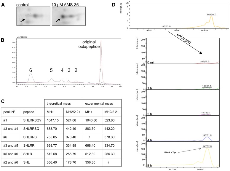
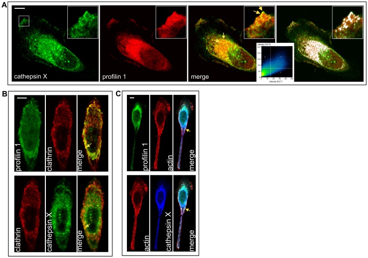
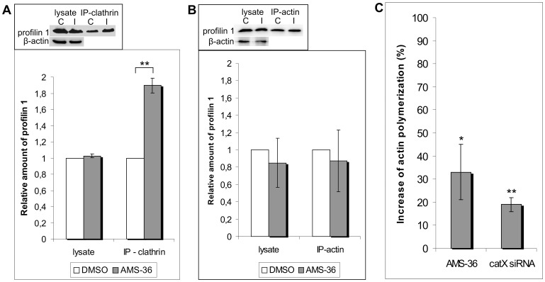
Cathepsin X has been reported to be a tumor promotion factor in various types of cancer; however, the molecular mechanisms linking its activity with malignant processes are not understood. Here we present profilin 1, a known tumor suppressor, as a target for cathepsin X carboxypeptidase activity in prostate cancer PC-3 cells. Profilin 1 co-localizes strongly with cathepsin X intracellularly in the perinuclear area as well as at the plasma membrane. Selective cleavage of C-terminal amino acids was demonstrated on a synthetic octapeptide representing the profilin C-terminal region, and on recombinant profilin 1. Further, intact profilin 1 binds its poly-L-proline ligand clathrin significantly better than it does the truncated one, as shown using cathepsin X specific inhibitor AMS-36 and immunoprecipitation of the profilin 1/clathrin complex. Moreover, the polymerization of actin, which depends also on the binding of poly-L-proline ligands to profilin 1, was promoted by AMS-36 treatment of cells and by siRNA cathepsin X silencing. Our results demonstrate that increased adhesion, migration and invasiveness of tumor cells depend on the inactivation of the tumor suppressive function of profilin 1 by cathepsin X. The latter is thus designated as a target for development of new antitumor strategies.


Conflict of interest statement

Competing Interests: The authors have declared that no competing interests exist.

Figures

Figure 1
Figure 1. Cathepsin X in MCF-7, MDA-MB-231 and PC-3 cells.
(A) Activity was measured in cell lysates using cathepsin X specific substrate Abz-FEK(Dnp)-OH. Mean values of 5 independent experiments are shown. ***P<0,001 (B) The amount of cathepsin X (ng/ml) in different cell lines was determined with ELISA. Mean values of 2 (MCF-7 and MDA-MB-231) or 4 (PC-3) independent experiments are shown.
Figure 2
Figure 2. Cathepsin X increases migration, adhesion and invasion of cancer cells.
Migration (A, B), adhesion (C) and invasion (D) assays were done using xCELLigence System. (A and B) Diagrams show a slope (cell index vs. time) of cells that migrated from the upper to the lower chamber. Cells migrated in the presence of DMSO (red line on graph) or 10 µM inhibitor of cathepsin X (green line on graph) (A) or cells, transfected with control (red line on graph) or cathepsin X specific siRNA (green line on graph) were used (B). (C) Diagram shows a slope for cells transfected with a control (red line on graph) or cathepsin X specific siRNA (green line on graph), that adhered to fibronectin (10 µg/ml). (D) Diagram shows a slope for cells transfected with a control (red line on graph) or cathepsin X specific siRNA (green line on graph) that invaded through Matrigel from the upper to the lower chamber. ***P≤0.01; ***P<0,001. Graphs show real-time curves of cell index (CI) as a function of time. Vertical lines represent the start and end of time intervals within which corresponding diagrams are calculated. Four (A), eight (B), four (C) and five (D) biological repeats were performed.
Figure 3
Figure 3. Identification of profilin 1 as a substrate for cathepsin X carboxypeptidase activity.
(A) Control versus AMS-36 treated sample is shown after 2D electrophoresis. The spot marked with arrow was identified as human profilin 1. (B and C) The C-terminal of profilin 1 (SHLRRSQY) (800 µM) was digested with recombinant cathepsin X (4.62 µM) at 37°C for 30 minutes and separated on a C18 Gemini column (5 µm, 110 Å, 150×4.6 mm) (Phenomenex). (B) 5 additional peaks, named peaks 2 to 6, were detected besides the original octapeptide (black line). The octapeptide control without enzyme is shown in red. (C) Q-TOF Premier mass spectrometry analysis of each peak showed the presence of 3 to 7 amino acid long peptides, all shortened by 1 amino acid from the C- terminal. (D) Profilin 1 (1 µg/µl; Abcam) was digested with recombinant cathepsin X (46.2 µM) at 37°C for several hours and the digestion product detected with mass spectrometry. A new peak was detected with molecular mass matching the mass of profilin 1 without the last amino acid residue Tyr.
Figure 4
Figure 4. Co-localization of cathepsin X and profilin 1 (A) with clathrin (B) and actin (C).
All proteins were visualized by immunofluorescence staining using primary antibodies to cathepsin X, profilin 1 and clathrin, followed by Alexa Fluor conjugated secondary antibodies Alexa Fluor 488, 555 and 633 or phalloidin conjugate for actin. (A) Cathepsin X is shown in green, profilin 1 in red and co-localization in yellow. For co-localization, also Zen 2011 Software (Carl Zeiss) option for improved visibility of co-localized pixels was used and co-localization is shown in white with corresponding scatter diagram. (B) Clathrin is shown in red, profilin 1 and cathepsin X are both in green due to clearer merged image. (C) Profilin 1 is shown in green, cathepsin X in blue and actin in red. Zen 2011 Software option for improved visibility of co-localization is used. Bars, 10 µm.
Figure 5
Figure 5. Cathepsin X modulates profilin 1 function by affecting the binding of poly-L-proline ligands.
(A and B) Profilin 1 forms a stable complex with actin and clathrin. Representative co-immunoprecipitations of profilin 1 with clathrin (A-inset) and actin (B-inset) in PC-3 cells, treated with DMSO or cathepsin X specific inhibitor. (A and B insets) Cell lysates were treated with anti-clathrin or anti β-actin antibody and immunoprecipitated on Protein A Sepharose beads. Profilin 1 was detected by Western blot in total cell lysates and in immunoprecipitated pellets. β-actin was used as a loading control. (A and B) Quantification of data from the insets. The graphs represent densitometric analysis of bands using Sygene's GeneTools Software (Sygene, U.K.). Three or two biological experiments indicate the normalized amount of profilin 1 that is in complex with clathrin (A) or actin (B), respectively. **P≤0.01. (C) Cathepsin X regulates actin polymerization. Flow cytometric analysis of permeabilized PC-3 cells is shown. Filamentous actin was stained with phalloidin conjugate. Cells were treated with DMSO or AMS-36 or were transfected with control or cathepsin X specific siRNA. Increase in actin polymerization is shown with values of both control experiments set to 0% increase in actin polymerization. Values are representative of four independent experiments. *P≤0.05; **P<0.01.

Similar articles

Cited by

References

    1. Turk V, Kos J, Turk B (2004) Cysteine cathepsins (proteases)–on the main stage of cancer? Cancer Cell 5: 409–410. - PubMed
    1. Nägler DK, Krüger S, Kellner A, Ziomek E, Menard R, et al. (2004) Up-regulation of cathepsin X in prostate cancer and prostatic intraepithelial neoplasia. Prostate 60: 109–119. - PubMed
    1. Fröhlich E, Schlagenhauff B, Möhrle M, Weber E, Klessen C, et al. (2001) Activity, expression, and transcription rate of the cathepsins B, D, H, and L in cutaneous malignant melanoma. Cancer 91: 972–982. - PubMed
    1. Krueger S, Kalinski T, Hundertmark T, Wex T, Küster D, et al. (2005) Up-regulation of cathepsin X in Helicobacter pylori gastritis and gastric cancer. J Pathol 207: 32–42. - PubMed
    1. Wang J, Chen L, Li Y, Guan XY (2011) Overexpression of cathepsin Z contributes to tumor metastasis by inducing epithelial-mesenchymal transition in hepatocellular carcinoma. PLoS One 6: e24967. - PMC - PubMed

Publication types

MeSH terms