. 2013;7(1):e1988.
doi: 10.1371/journal.pntd.0001988. Epub 2013 Jan 10.

Functional mapping of protein kinase A reveals its importance in adult Schistosoma mansoni motor activity

Affiliations

Functional mapping of protein kinase A reveals its importance in adult Schistosoma mansoni motor activity

Paulu S R de Saram et al. PLoS Negl Trop Dis. 2013.

Abstract

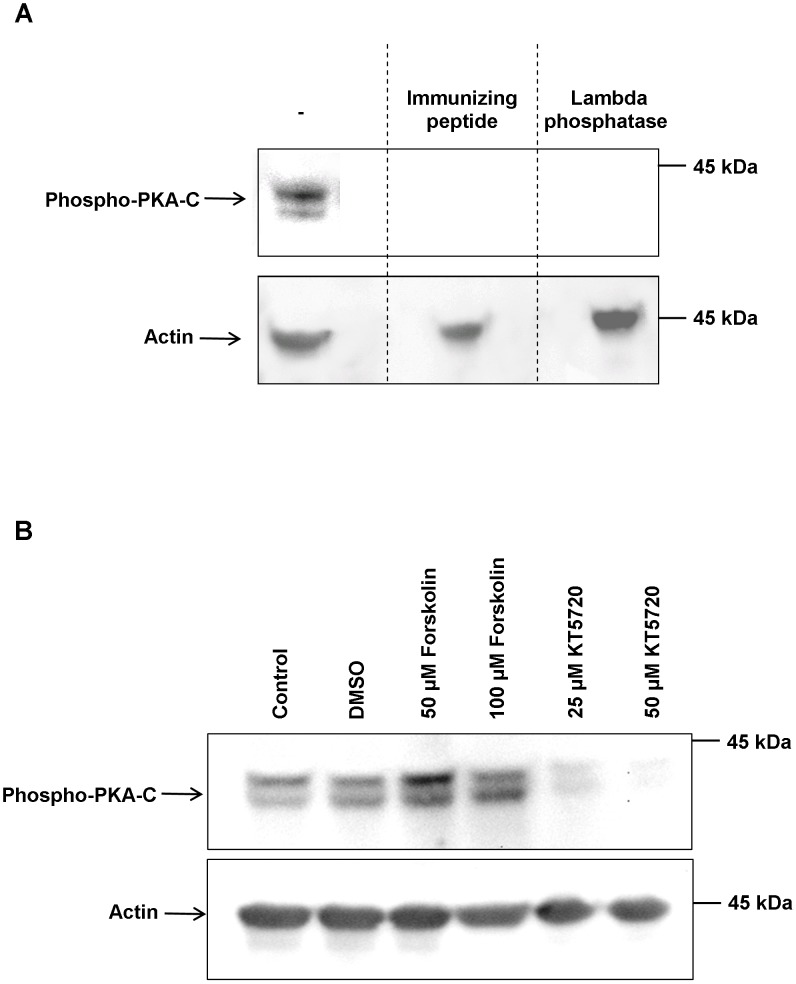
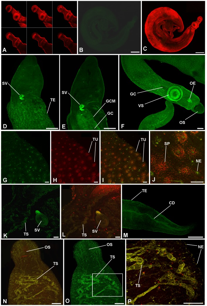
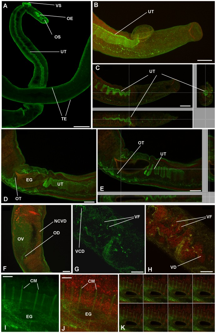
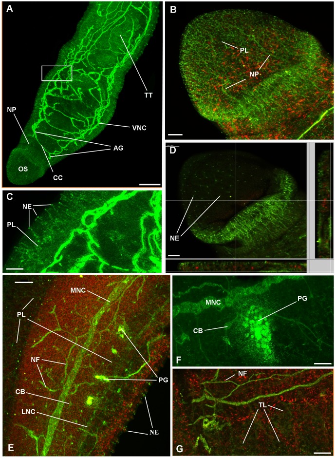
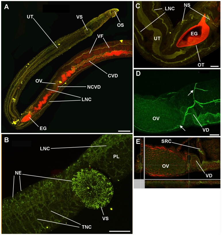
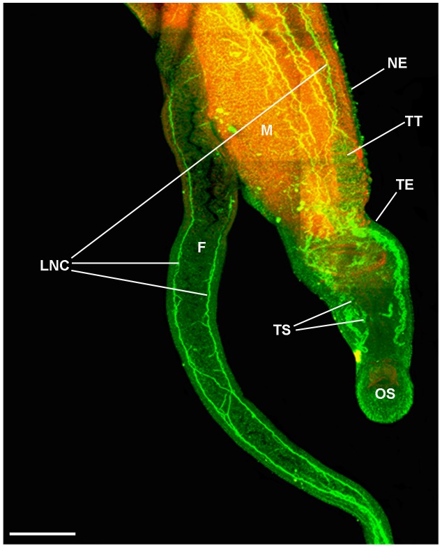
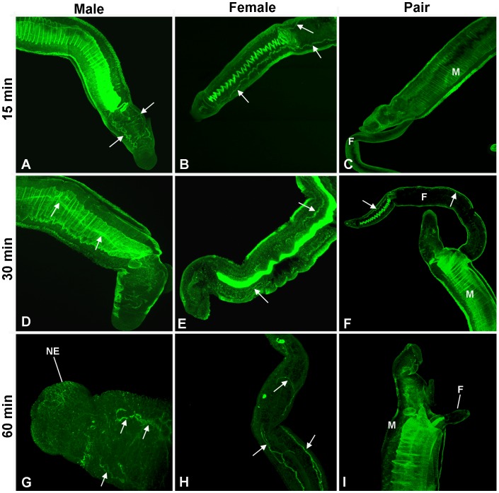
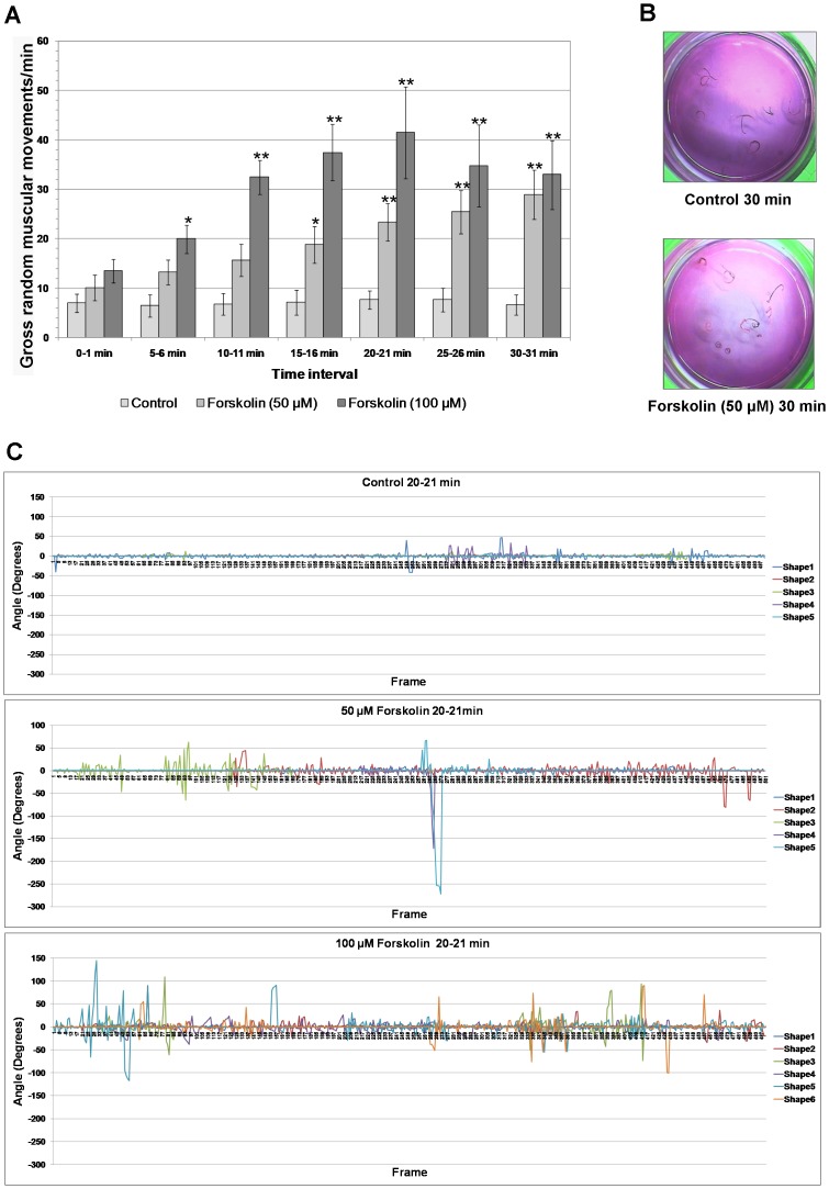
Cyclic AMP (cAMP)-dependent protein kinase/protein kinase A (PKA) is the major transducer of cAMP signalling in eukaryotic cells. Here, using laser scanning confocal microscopy and 'smart' anti-phospho PKA antibodies that exclusively detect activated PKA, we provide a detailed in situ analysis of PKA signalling in intact adult Schistosoma mansoni, a causative agent of debilitating human intestinal schistosomiasis. In both adult male and female worms, activated PKA was consistently found associated with the tegument, oral and ventral suckers, oesophagus and somatic musculature. In addition, the seminal vesicle and gynaecophoric canal muscles of the male displayed activated PKA whereas in female worms activated PKA localized to the ootype wall, the ovary, and the uterus particularly around eggs during expulsion. Exposure of live worms to the PKA activator forskolin (50 µM) resulted in striking PKA activation in the central and peripheral nervous system including at nerve endings at/near the tegument surface. Such neuronal PKA activation was also observed without forskolin treatment, but only in a single batch of worms. In addition, PKA activation within the central and peripheral nervous systems visibly increased within 15 min of worm-pair separation when compared to that observed in closely coupled worm pairs. Finally, exposure of adult worms to forskolin induced hyperkinesias in a time and dose dependent manner with 100 µM forskolin significantly increasing the frequency of gross worm movements to 5.3 times that of control worms (P≤0.001). Collectively these data are consistent with PKA playing a central part in motor activity and neuronal communication, and possibly interplay between these two systems in S. mansoni. This study, the first to localize a protein kinase when exclusively in an activated state in adult S. mansoni, provides valuable insight into the intricacies of functional protein kinase signalling in the context of whole schistosome physiology.


Conflict of interest statement

The authors have declared that no competing interests exist.

Figures

Figure 1
Figure 1. Western blot analysis demonstrating antibody specificity and activation and inhibition of S. mamsoni PKA-C.
(A) Adult S. mansoni protein extracts (from one worm pair) were processed for western blotting with anti-phospho-PKA-C antibodies as described in Materials and Methods; the immunizing peptide and lambda phosphatase were employed to confirm that the antibody reacted with phosphorylated PKA-C and not the non-phosphorylated form. (B) Live adult S. mansoni were incubated in 50 µM or 100 µM forskolin, 25 µM or 50 µM KT5720, vehicle (DMSO), or DMEM alone (Control) for 1 h at 28°C and worm protein extracts processed for western blotting with anti-phospho-PKA-C antibodies. Anti-actin antibodies were used to assess any differences in protein loading between samples. The results shown in each panel are representative of data from two independent experiments.
Figure 2
Figure 2. In situ distribution of phosphorylated (activated) PKA (green) in adult male S. mansoni revealed using anti-phospho-PKA-C antibodies.
Schistosomes were fixed and stained as detailed in Materials and Methods. Images show z-axis projections in maximum pixel brightness mode except (A) which shows selected serial optical z-sections; images I, J, L, N, and P are overlays of activated PKA with F-actin stained by rhodamine phalloidin (red). (A–C) Negative control worms incubated without primary antibodies but with Alexa Fluor 488 secondary antibodies and rhodamine phalloidin. In male worms, prominent PKA activation is particularly associated with: (D, anterior dorsal surface scan) the tegument (TE); (E, deeper body scan) gynaecophoric canal muscles (GCM) of the gynaecophoric canal (GC), and seminal vesicle (SV); (F, anterior ventral surface scan) ventral sucker (VS), oral sucker (OS) and oesophagus (OE); (G–J) tubercles (TU) of the tegument, areas proximal to the centres of the tubercle spines (SP) and at surface-proximal nerve endings (NE) are also evident in (P); (K, L, N–P) tubular structures (TS) of unknown function close to the SV (K, L) also in the anterior of the worm (N–P, with boxed region shown in O, enlarged for clarity in P); (M) posterior region displaying activated PKA associated with the collecting duct (CD). Bar: B, C = 300 µm; D–F, M = 100 µm; G–L, P = 20 µm; N,O = 50 µm.
Figure 3
Figure 3. In situ distribution of phosphorylated (activated) PKA (green) in adult female S. mansoni revealed using anti-phospho-PKA-C antibodies.
Schistosomes were fixed and stained as detailed in Materials and Methods. Images show z-axis projections in maximum pixel brightness mode with further z, x and y, z projections in C and E; K shows serial optical z-sections; images B–F, H, J and K are overlays of activated PKA with F-actin stained by rhodamine phalloidin (red). In female worms, prominent PKA activation is particularly associated with: (A) the tegument (TE), oesophagus (OE), ventral and oral suckers (VS/OS); (A–E) uterus (UT); (D, E) ootype (OT) surrounding the egg (EG) which also autofluoresces red in D and E; (F) ovary (OV) and oviduct (OD) but not the non-ciliated vitelline duct (NCVD); (G, H) vitelline collective duct (VCD) structures and areas of the vitelline follicles (VF) leading to the vitelline duct (VD); (I–K, taken from female in copula) circular muscles (CM) revealed as ring-like distributions of activated PKA during egg (EG) expulsion along the uterus. Bar: A = 100 µm; B–J = 40 µm.
Figure 4
Figure 4. Activated PKA associates with the nervous system of adult male S. mansoni.
Schistosomes were fixed and stained with anti-phospho-PKA-C antibodies (green) and rhodamine phalloidin (red) to localize activated PKA and actin, respectively, as detailed in Materials and Methods. Images show z-axis projections in maximum pixel brightness mode with further z, x and y, z projections in D. Intense PKA activation is particularly associated with: (A, anterior view) ventral nerve cords (VNC), connecting cerebral commissures (CC), and anterior ganglia (AG); (A, B) complex nerve plexus (PL) associated with the oral sucker (OS) and extending nerve processes (NP); (C, enlarged from boxed region shown in A) and (D) nerve endings (NE) emanating from the PL evident over the body surface including on the tegument and oral sucker; (E, F) peripheral nervous system including PL extending from the lateral nerve cord (LNC), main nerve cord (MNC), complex PL extending to the underlying musculature (red), surface proximal NEs extending from the PL, nerve fibres (NF); peripheral ganglia (PG) and cell bodies (CB) which connect with the MNC; (F) the ganglion shown was close to the surface of the gut. Additionally, diffuse PKA activation is associated with the (A, G) testicular lobes (TL) of the testes (TT), which are surrounded by the testicular capsules stained red. Bar: A = 100 µm; B–D, F, G = 20 µm; E = 40 µm.
Figure 5
Figure 5. Activated PKA associates with the nervous system of adult female S. mansoni.
Schistosomes were fixed and stained with anti-phospho-PKA-C antibodies (green) and rhodamine phalloidin (red) to localize activated PKA and actin, respectively, as detailed in Materials and Methods. Images show z-axis projections in maximum pixel brightness mode with further z, x and y, z projections in E. Activated PKA is particularly associated with: (A) longitudinal nerve cord (LNC), neural structures in the oral sucker (OS), ciliated vitelline duct (CVD) adjacent to the vitelline follicles (VF) but not the non-ciliated vitelline duct (NCVD); (B) neural structures of the ventral sucker (VS), surface-proximal nerve endings (NE) extending from the complex nerve plexus (PL) including those on the VS, and transverse nerve cords (TNC); (C) neural structures (NS) innervating the ootype (OT) surrounding the egg (EG), LNC and uterus (UT); (D and E) ovary (OV) and innervating nerves (arrowed) which extend to regions including the vitelline ducts (VD, obscuring the oviducts) and the seminal receptacle complex (SRC). Bar: A = 100 µm; B, D = 40 µm; C = 20 µm.
Figure 6
Figure 6. Immunolocalization of activated PKA in forskolin-treated adult male (M) and female (F) S. mansoni in copula.
Worm couples were incubated in 50 µM forskolin for 1 h and fixed and stained with anti-phospho-PKA-C antibodies (green) and rhodamine phalloidin (red) to localize activated PKA, and actin, respectively, as detailed in Materials and Methods. Intense PKA activation is seen in the tegument (TE) musculature and the nervous system including the longitudinal nerve cords (LNC) and nerve endings (NE) covering the TE and oral sucker (OS). The uncharacterized tubular structures (TS) previously show in Figure 2 also display activated PKA. Image shows a z-axis projection in maximum pixel brightness mode. Bar = 100 µm.
Figure 7
Figure 7. Activation of PKA in the nervous system of adult male (M) and female (F) S. mansoni upon pair separation.
Schistosomes were fixed (A–C) 15 min, (D–F) 30 min, or (G–I) 60 min after separation and were stained with anti-phospho-PKA-C antibodies (green) to localize activated PKA as detailed in Materials and Methods; schistosomes that remained in copula (pair; C,F,I) for these durations were also processed for comparison with separated males (A,D,G) or females (B,E,H). Images show z-axis projections in maximum pixel brightness mode. The arrows highlight regions of PKA activation within the nervous system; the putative surface-proximal nerve endings (NE) are also indicated.
Figure 8
Figure 8. Forskolin increases the frequency and angle of S. mansoni adult worm movements.
(A) Adult worms were incubated in either 50 µM or 100 µM forskolin, or DMSO vehicle (control) for various durations (0–30 min) and were filmed for 1 min and analyzed as detailed in Materials and Methods. Values shown represent mean number of gross random muscular movements per minute (±SEM, n = 7 for each treatment at each time point); *P≤0.05; **P≤0.001. (B) An increased propensity of worm coiling was observed following incubation with 50 µM forskolin when compared to controls; effects after 30 min are illustrated. (C) Detailed analysis of individual worm (shape) movements using the ImageJ plugin wrMTrck when worms were incubated in either 50 µM or 100 µM forskolin, or DMSO vehicle (control) for 20 min. The angle (degrees) of worm movement is shown as a function of time (frame) during 1 min recording; results of at least 5 individual worms are shown.

Similar articles

Cited by

References

    1. Steinmann P, Keiser J, Bos R, Tanner M, Utzinger J (2006) Schistosomiasis and water resources development: systematic review, meta-analysis, and estimates of people at risk. Lancet Infect Dis 6: 411–425. - PubMed
    1. Walker AJ (2011) Insights into the functional biology of schistosomes. Parasite Vectors 4: 203. - PMC - PubMed
    1. Popiel I, Basch PF (1984) Reproductive development of female Schistosoma mansoni (Digenia: Schistosomatidae) following bisexual pairing of worms and worm segments. J Exp Zool 232: 141–150. - PubMed
    1. Kuntz W (2001) Schistosome male-female interaction: induction of germ-cell differentiation. Trends Parasitol 17: 227–231. - PubMed
    1. Gelanti SE, Huang SCC, Pearce EJ (2012) Cell death and reproductive regression in female Schistosoma mansoni . PLoS Negl Trop Dis 6: e1509. - PMC - PubMed

Publication types

Substances

LinkOut - more resources