

Outcomes after bronchoscopic procedures for primary tracheobronchial amyloidosis: retrospective study of 6 cases
- PMID: 23326661
- PMCID: PMC3544315
- DOI: 10.1155/2012/352719
Outcomes after bronchoscopic procedures for primary tracheobronchial amyloidosis: retrospective study of 6 cases
Abstract
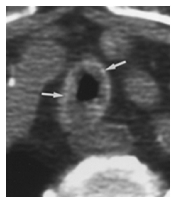
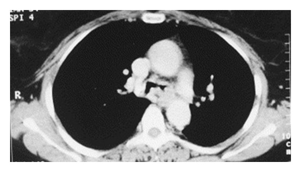
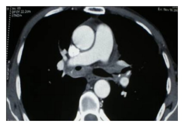
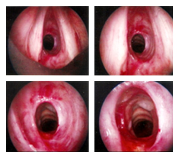
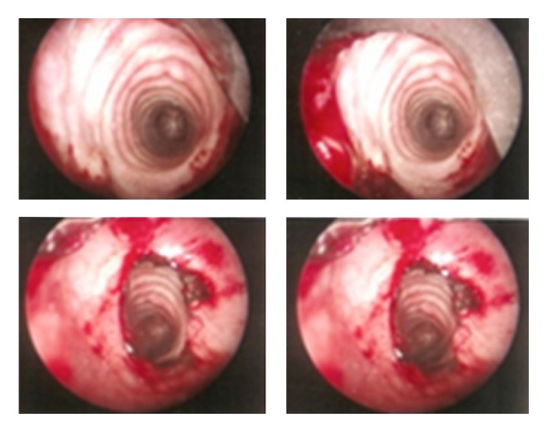
Respiratory amyloidosis is a rare disease which refers to localized aberrant extracellular protein deposits within the airways. Tracheobronchial amyloidosis (TBA) refers to the deposition of localized amyloid deposits within the upper airways. Treatments have historically focused on bronchoscopic techniques including debridement, laser ablation, balloon dilation, and stent placement. We present the outcomes after rigid bronchoscopy to remove the amyloid protein causing the airway obstruction in 6 cases of tracheobronchial amyloidosis. This is the first report of primary diffuse tracheobronchial amyloidosis in our department; clinical features, in addition to therapy in the treatment of TBA, are reviewed. This paper shows that, in patients with TBA causing airway obstruction, excellent results can be obtained with rigid bronchoscopy and stenting of the obstructing lesion.
Figures
Similar articles
-
Tracheobronchial amyloidosis. The Boston University experience from 1984 to 1999.Medicine (Baltimore). 2000 Mar;79(2):69-79. doi: 10.1097/00005792-200003000-00001. Medicine (Baltimore). 2000. PMID: 10771705
-
Tracheobronchial amyloidosis treated with rigid bronchoscopy and stenting.Surg Endosc. 2003 Apr;17(4):658-9. doi: 10.1007/s00464-002-4260-z. Epub 2003 Feb 10. Surg Endosc. 2003. PMID: 12574930
-
Tracheobronchial amyloidosis: a case report of successful treatment with external beam radiation therapy.Chest. 2004 Feb;125(2):784-9. doi: 10.1378/chest.125.2.784. Chest. 2004. PMID: 14769766
-
A case of primary diffuse tracheobronchial amyloidosis.Ann Thorac Surg. 2004 May;77(5):1832-4. doi: 10.1016/S0003-4975(03)00999-8. Ann Thorac Surg. 2004. PMID: 15111203 Review.
-
Primary localized tracheobronchial amyloidosis presenting with massive hemoptysis: a case report and literature review.Clin Respir J. 2017 Jan;11(1):122-125. doi: 10.1111/crj.12301. Epub 2015 Apr 28. Clin Respir J. 2017. PMID: 25832552 Review.
Cited by
-
An unusual case of hoarseness of voice.Respir Med Case Rep. 2018 Feb 2;23:128-130. doi: 10.1016/j.rmcr.2018.01.013. eCollection 2018. Respir Med Case Rep. 2018. PMID: 29719798 Free PMC article.
-
Silicone stent placement for primary tracheal amyloidosis accompanied by cartilage destruction.Tuberc Respir Dis (Seoul). 2014 Jun;76(6):292-4. doi: 10.4046/trd.2014.76.6.292. Epub 2014 Jun 28. Tuberc Respir Dis (Seoul). 2014. PMID: 25024724 Free PMC article.
-
A rare cause of stridor: isolated tracheal amyloidosis.Can Respir J. 2014 Sep-Oct;21(5):273-5. doi: 10.1155/2014/174092. Can Respir J. 2014. PMID: 25299219 Free PMC article.
-
Multifocal primary amyloidosis of the airways: Case report and review of the literature.Respir Med Case Rep. 2015 May 27;15:115-7. doi: 10.1016/j.rmcr.2015.05.004. eCollection 2015. Respir Med Case Rep. 2015. PMID: 26236619 Free PMC article.
-
Tracheobronchial amyloidosis: A case report and review of the literature.J Case Rep Med. 2014;3:235859. doi: 10.4303/jcrm/235859. J Case Rep Med. 2014. PMID: 26998363 Free PMC article.
References
-
- Capizzi SA, Betancourt E, Prakash UBS. Tracheobronchial amyloidosis. Mayo Clinic Proceedings. 2000;75(11):1148–1152. - PubMed
-
- Howard ME, Ireton J, Daniels F, Langton D, Manolitsas ND, Fogarty P. Pulmonary presentations of amyloidosis. Respirology. 2001;6:61–64. - PubMed
-
- Paccalin M, Hachulla E, Cazalet C, et al. Localized amyloidosis: a survey of 35 French cases. Amyloid. 2005;12(4):239–245. - PubMed
-
- Gibbaoui H, Abouchacra S, Yaman M. A case of primary diffuse tracheobronchial amyloidosis. Annals of Thoracic Surgery. 2004;77(5):1832–1834. - PubMed
LinkOut - more resources
Full Text Sources
