

Imaging findings of pelvic tumor thrombosis extending from sacral bone metastasis of adrenocortical carcinoma
- PMID: 23326744
- PMCID: PMC3541597
- DOI: 10.1155/2012/919603
Imaging findings of pelvic tumor thrombosis extending from sacral bone metastasis of adrenocortical carcinoma
Abstract
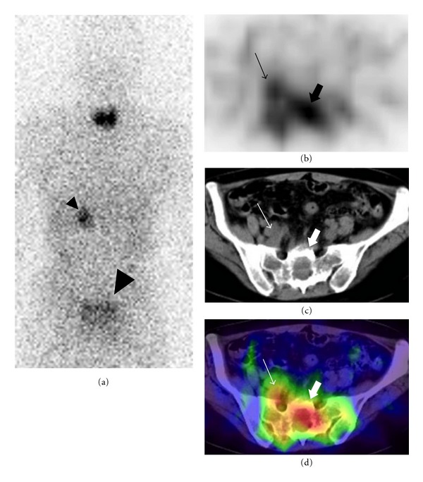
We report the imaging findings of a patient with adrenocortical carcinoma who showed pelvic tumor thrombosis extending from sacral bone metastasis. Contrast-enhanced computed tomography demonstrated extensive intraluminal filling defects in the pelvic veins. A lytic lesion in the sacrum was also noted and continuity between the sacral lesion and the filling defect in the branch of pelvic veins was indicated. The filling defects showed increased uptake on positron emission tomography with (18)F-fluorodeoxyglucose and single-photon emission computed tomography with (131)I-iodomethylnorcholesterol, and fusion images with computed tomography aided the localization of the increased uptake areas. Multimodality imaging may be beneficial for the characterization and localization of lesions in patients suspected of having metastatic adrenocortical carcinoma.
Figures


References
-
- Figueroa AJ, Stein JP, Lieskovsky G, Skinner DG. Adrenal cortical carcinoma associated with venous tumour thrombus extension. British Journal of Urology. 1997;80(3):397–400. - PubMed
-
- Senthil R, Mittal BR, Kashyap R, Bhattacharya A, Radotra BD, Bhansali A. 18F FDG PET/CT demonstration of IVC and right atrial involvement in adrenocortical carcinoma. Japanese Journal of Radiology. 2012;30:281–283. - PubMed
-
- Yavascaoglu I, Yilmaz M, Kordan Y. Cardiac and caval invasion of left adrenocortical carcinoma. Urologia Internationalis. 2008;81(2):244–246. - PubMed
-
- Bhargava P, Kumar R, Zhuang H, Charron M, Alavi A. Catheter-Related Focal FDG Activity on Whole Body PET Imaging. Clinical Nuclear Medicine. 2004;29(4):238–242. - PubMed
-
- Sharma P, Kumar R, Jeph S, Karunanithi S, Naswa N, Gupta A, et al. 18F-FDG PET-CT in the diagnosis of tumor thrombus: can it be differentiated from benign thrombus? Nuclear Medicine Communications. 2011;32:782–788. - PubMed
LinkOut - more resources
Full Text Sources
