Determination and comparison of specifics of nucleus pulposus cells of human intervertebral disc in alginate and chitosan-gelatin scaffolds
- PMID: 23326811
- PMCID: PMC3544085
- DOI: 10.4103/2277-9175.102996
Determination and comparison of specifics of nucleus pulposus cells of human intervertebral disc in alginate and chitosan-gelatin scaffolds
Abstract
Introduction: Low back pain is a major economical and social problem nowadays. Intervertebral disc herniation and central degeneration of disc are two major reasons of low back pain that occur because of structural impairment of disc. The intervertebral disc contains three parts as follows : Annulus fibrosus, transitional region, and nucleus pulposus, which forms the central nucleus of the disc. The reduction of cell count and extracellular matrix, especially in nucleus pulposus, causes disc degeneration. Different scaffolds (natural and synthetic) have been used for tissue repairing and regeneration of the intervertebral disc in tissue engineering. Most scaffolds have biodegradable and biocompatible characteristics and also prepare a fine condition for proliferation and migration of cells. In this study, proliferation of NP cells of human intervertebral disc compromised in Chitosan-gelatin scaffold with alginate scaffold was studied.
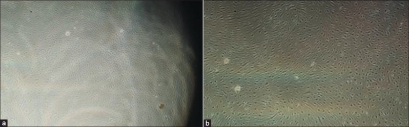
Materials and methods: NP cells derived from nucleus pulposus by collagenase enzymatic hydrolysis. They were derived from patients who undergoing open surgery for discectomy in the Isfahan Alzahra hospital. Chitosan was blended with gelatin and glutaraldehyde was used for cross linking the two polymers. Then, alginate scaffold was prepared. Cellular suspension with 1 × 10(5) transferred to each scaffold and cultured for 21 days. Cell viability and proliferation investigated by trypan blue and (3-(4,5-Dimethylthiazol-2-yl)-2,5-diphenyltetrazolium bromide (MTT) assay. Scanning electron microscope (SEM) was used to assert the porosity and to survey structure of scaffold.
Results: MTT assay dem1onstrated that cell viability of third day had significant difference in contrast by first day in both scaffolds. Accordingly, there was a significant decreased in cellular viability from day 3 to 21. Results of the cell count showed a punctual elevation cell numbers for alginate scaffold but there was no similar result for chitosan-gelatin scaffold.
Conclusion: Alginate scaffold prepared a better condition for proliferation of NP cells in comparison with chitosan-gelatin scaffold. Results of this study suggest that alginate scaffold could be useful in in vivo studies and treatment.
Keywords: Alginate scaffold; NP cells; chitosan-gelatin scaffold; intervertebral disc; tissue enginiaring.
Conflict of interest statement
Figures


Similar articles
-
Evaluation of the proliferation and viability rates of nucleus pulposus cells of human intervertebral disk in fabricated chitosan-gelatin scaffolds by freeze drying and freeze gelation methods.Adv Biomed Res. 2015 Nov 30;4:251. doi: 10.4103/2277-9175.170676. eCollection 2015. Adv Biomed Res. 2015. PMID: 26918233 Free PMC article.
-
Enhancing cell migration in shape-memory alginate-collagen composite scaffolds: In vitro and ex vivo assessment for intervertebral disc repair.J Biomater Appl. 2015 Apr;29(9):1230-46. doi: 10.1177/0885328214557905. Epub 2014 Nov 6. J Biomater Appl. 2015. PMID: 25376622
-
[Fabrication and analysis of a novel tissue engineered composite biphasic scaffold for annulus fibrosus and nucleus pulposus].Zhongguo Xiu Fu Chong Jian Wai Ke Za Zhi. 2013 Apr;27(4):475-80. Zhongguo Xiu Fu Chong Jian Wai Ke Za Zhi. 2013. PMID: 23757878 Chinese.
-
Repair and Regenerative Therapies of the Annulus Fibrosus of the Intervertebral Disc.J Coll Physicians Surg Pak. 2016 Feb;26(2):138-44. J Coll Physicians Surg Pak. 2016. PMID: 26876403 Review.
-
Treatment of the degenerated intervertebral disc; closure, repair and regeneration of the annulus fibrosus.J Tissue Eng Regen Med. 2015 Oct;9(10):1120-32. doi: 10.1002/term.1866. Epub 2014 Feb 25. J Tissue Eng Regen Med. 2015. PMID: 24616324 Review.
Cited by
-
Confocal scanning of intervertebral disc cells in 3D: Inside alginate beads and in native microenvironment.JOR Spine. 2020 Jul 15;3(4):e1106. doi: 10.1002/jsp2.1106. eCollection 2020 Dec. JOR Spine. 2020. PMID: 33392446 Free PMC article.
-
Multiscale Regulation of the Intervertebral Disc: Achievements in Experimental, In Silico, and Regenerative Research.Int J Mol Sci. 2021 Jan 12;22(2):703. doi: 10.3390/ijms22020703. Int J Mol Sci. 2021. PMID: 33445782 Free PMC article. Review.
-
Evaluation of the proliferation and viability rates of nucleus pulposus cells of human intervertebral disk in fabricated chitosan-gelatin scaffolds by freeze drying and freeze gelation methods.Adv Biomed Res. 2015 Nov 30;4:251. doi: 10.4103/2277-9175.170676. eCollection 2015. Adv Biomed Res. 2015. PMID: 26918233 Free PMC article.
-
Cationic antimicrobial polymers and their assemblies.Int J Mol Sci. 2013 May 10;14(5):9906-46. doi: 10.3390/ijms14059906. Int J Mol Sci. 2013. PMID: 23665898 Free PMC article. Review.
-
Injectable hydrogel provides growth-permissive environment for human nucleus pulposus cells.J Biomed Mater Res A. 2016 Feb;104(2):419-26. doi: 10.1002/jbm.a.35580. Epub 2015 Oct 15. J Biomed Mater Res A. 2016. PMID: 26422588 Free PMC article.
References
-
- Waddell G. Low back pain: A twentieth century health care enigma. Spine. 1966;21:2820–5. - PubMed
-
- Mauth C, Bono E, Haas S, Paesold G, Wiese H, Maier G, et al. Cell-seeded polyurethane-fibrin structures-a possible system for intervertebral disc regeneration. Eur Cell Mater. 2009;18:27–39. - PubMed
-
- Boss N, Rieder R, Schade V, Spartt KF, Semmer N, Aebim The diagnostic accuracy of magnetic resonance imaging, work perception, and psychosocial factors in identifying symptomatic descherniations. Spine. 1995;20:2613–25. - PubMed
-
- Oegema TR., Jr The role of disc cell heterogeneity in determining disc biochemistry: A speculation. Biochem Soc Trans. 2002;30:839–44. - PubMed
LinkOut - more resources
Full Text Sources
Miscellaneous