. 2013 Jan 17:8:1.
doi: 10.1186/1749-8104-8-1.

Proliferation zones in the axolotl brain and regeneration of the telencephalon

Affiliations

Proliferation zones in the axolotl brain and regeneration of the telencephalon

Malcolm Maden et al. Neural Dev. .

Abstract

Background: Although the brains of lower vertebrates are known to exhibit somewhat limited regeneration after incisional or stab wounds, the Urodele brain exhibits extensive regeneration after massive tissue removal. Discovering whether and how neural progenitor cells that reside in the ventricular zones of Urodeles proliferate to mediate tissue repair in response to injury may produce novel leads for regenerative strategies. Here we show that endogenous neural progenitor cells resident to the ventricular zone of Urodeles spontaneously proliferate, producing progeny that migrate throughout the telencephalon before terminally differentiating into neurons. These progenitor cells appear to be responsible for telencephalon regeneration after tissue removal and their activity may be up-regulated by injury through an olfactory cue.

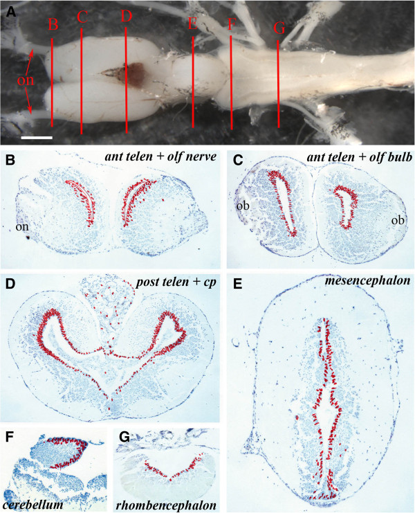
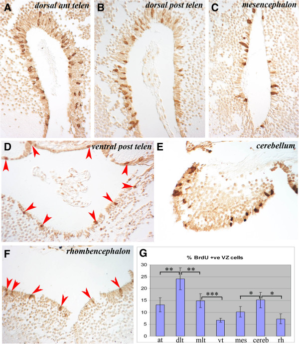
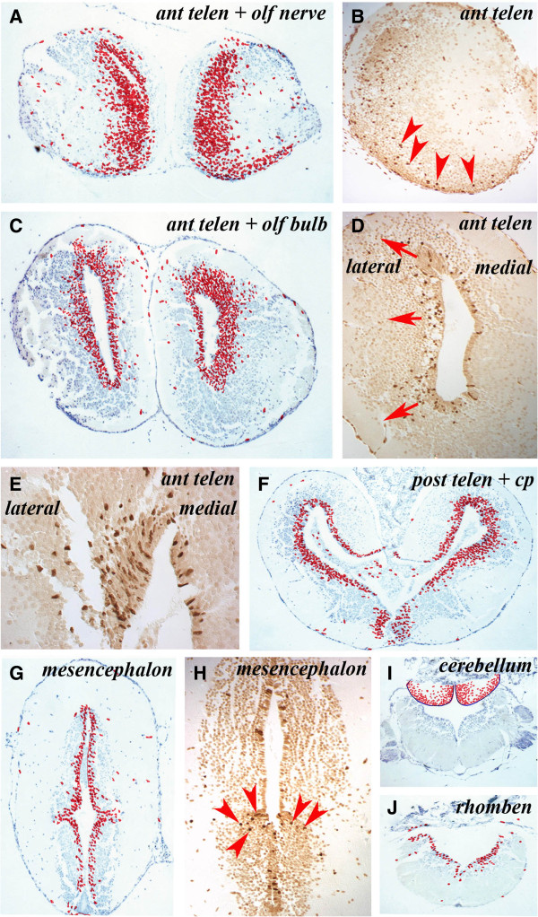
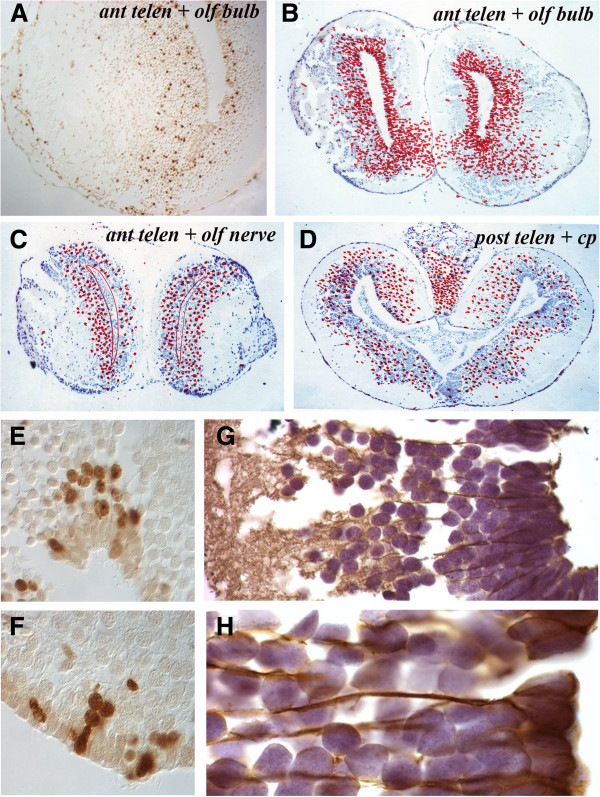
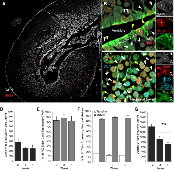
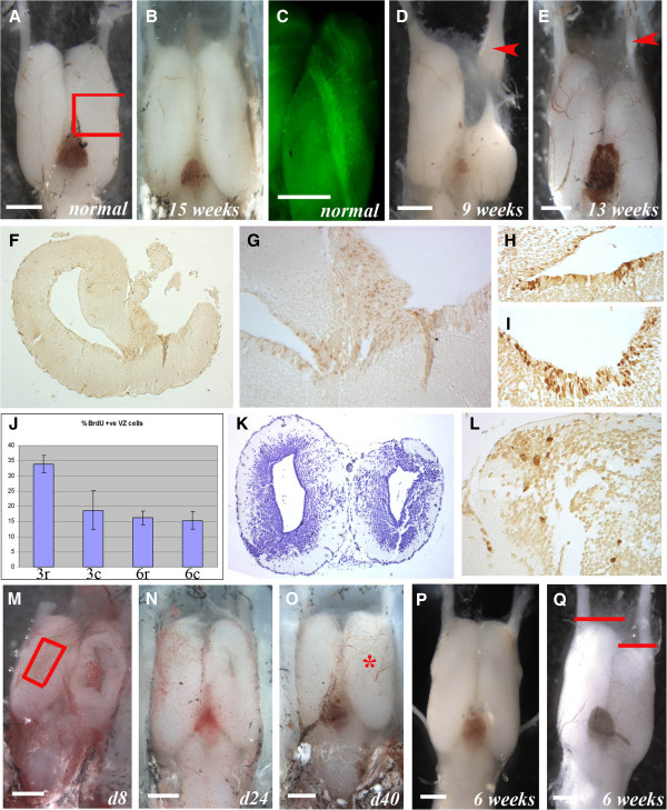
Results: There is extensive proliferation of endogenous neural progenitor cells throughout the ventricular zone of the adult axolotl brain. The highest levels are observed in the telencephalon, especially the dorsolateral aspect, and cerebellum. Lower levels are observed in the mesencephalon and rhombencephalon. New cells produced in the ventricular zone migrate laterally, dorsally and ventrally into the surrounding neuronal layer. After migrating from the ventricular zone, the new cells primarily express markers of neuronal differentiative fates. Large-scale telencephalic tissue removal stimulates progenitor cell proliferation in the ventricular zone of the damaged region, followed by proliferation in the tissue that surrounds the healing edges of the wound until the telencephalon has completed regeneration. The proliferative stimulus appears to reside in the olfactory system, because telencephalic regeneration does not occur in the brains of olfactory bulbectomized animals in which the damaged neural tissue simply heals over.

Conclusion: There is a continual generation of neuronal cells from neural progenitor cells located within the ventricular zone of the axolotl brain. Variable rates of proliferation were detected across brain regions. These neural progenitor cells appear to mediate telencephalic tissue regeneration through an injury-induced olfactory cue. Identification of this cue is our future goal.


Figures

Figure 1
Figure 1
Ventricular zones with variable proliferative activity can be detected throughout the axolotl central nervous system. (A) Structure of the axolotl brain showing the levels at which sections ventricular zone proliferation was analyzed. B = anterior telencephalon and olfactory nerve, C = anterior telencephalon and olfactory bulb, D = posterior telencephalon, E = mesencephalon, F = cerebellum, G = rhombencephalon; on: olfactory nerve. Scale bar = 1 mm. (B G) Cumulative BrdU+ cell numbers on five sections from each level depicted in A on which individual labeled cells are marked with a red dot. Regions of the brain are marked.; on: olfactory nerve; ob: olfactory bulb; cp: choroid plexus.
Figure 2
Figure 2
Cells labeled with BrdU after an overnight incubation are largely retained in the ventricular zone throughout the neuraxis, which shows regional variation in proliferative activity. (A-F) Examples of BrdU+ cells in the VZ of different brain regions show variation in numbers of labeled cells. Regions of the brain are marked. In D and F, BrdU+ cells are marked with red arrowheads. (G) Counts of proliferating VZ cells in different regions of the brain expressed as a percentage of total VZ cells. at: anterior telencephalon; BrdU: bromodeoxyuridine; cereb: cerebellum; dlt: dorsolateral telencephalon; mes: mesencephalon; mlt: mediolateral telencephalon; rh: rhombencephalon; vt: ventral telencephalon; VZ: ventricular zone. * significant (P = 0.025), ** very significant (P = 0.006), *** extremely significant (P = 0.0004) by Student’s T-test.
Figure 3
Figure 3
Sections taken one week after overnight BrdU labeling show the migration of BrdU+ cells from the ventricular zone into the mantle zone at different rates at different levels of the central nervous system. (A, C, F, G, I, J) Positions of labeled cells on five cumulative sections at the levels marked on the figure. The migration of BrdU+ cells outwards at different rates can be appreciated by comparing A and C with G. (B, D, E, H) Labeled cells on individual sections.
Figure 4
Figure 4
Migration patterns and glial fibrillary acidic protein expression in the telencephalon. (A) Single section through the anterior telencephalon and olfactory bulb taken 4 weeks after overnight BrdU labeling shows a stream of cells moving laterally into the olfactory bulb. (B) Cumulative positions of BrdU+ cells in the anterior telencephalon and olfactory bulb level 4 weeks after overnight labeling. (C) Cumulative positions of BrdU+ cells in the anterior telencephalon and olfactory nerve level 4 weeks after overnight labeling showing a decreased number of BrdU+ cells in the VZ. (D) Cumulative positions of BrdU+ cells in the posterior telencephalon 4 weeks after overnight labeling showing a decreased number of BrdU+ cells in the VZ. (E,F) Patterns of BrdU+ cells in and adjacent to the VZ 3 weeks after overnight labeling. (G,H) Glial fibrillary acidic protein immunocytochemistry of the normal telencephalon at low power (G) and high power (H) to show the cytoplasmic extensions of radial glial cells. BrdU: bromodeoxyuridine; VZ: ventricular zone.
Figure 5
Figure 5
Two weeks after BrdU labeling, new cells have migrated and differentiated into neurons and a few new cells are retained in the ventricular zone and express GFAP. (A) Confocal image at 10× magnification shows that, in the uninjured axolotl telencephalon, most BrdU+ cells have migrated into the mantle zone 2 weeks after labeling. A few BrdU+ cells are retained in the VZ. (B,C) Confocal images taken at 40× objective (2.3× digital zoom) of sections stained with 4-6-diamidino-2-phenylindole (in white; B1 and C1) and antibodies against (B) BrdU (in red; B2) and GFAP (in green; B3) or (C) BrdU (in red; C2), DCX (in blue; C3), NeuN (in green; C3). The image in (B) shows that very few BrdU+ cells are retained in the VZ by 2 weeks after overnight BrdU labeling, but those that do remain and express GFAP may represent new radial glia-like cells. (C) by 2 weeks, all BrdU+ cells in the mantle zone express the mature neuronal marker NeuN (white and yellow arrows) and a small percentage of the NeuN+ neurons retain DCX expression (yellow arrows) and are likely transitioning into mature neurons. (D) The relatively small densities (versus F) of BrdU/GFAP+ cells retained in the VZ are resilient across weeks 2 to 4 after BrdU labeling. (E) Most BrdU+ VZ cells express GFAP. (F) All BrdU+ cells in the neuronal layer express NeuN and a few NeuN+ neurons retain DCX expression. These percentages are consistent across weeks 2 to 4 after BrdU labeling. (G) New neurons are either vulnerable to cell death or are still migrating because their densities significantly decrease between weeks 2 and 3 (**P <0.001). BrdU: bromodeoxyuridine; DCX: doublecortin; GFAP: glial fibrillary acidic protein; VZ: ventricular zone.
Figure 6
Figure 6
Regeneration of the axolotl telencephalon. (A) Normal forebrain (ablated region marked with red box). (B) 15 weeks later showing complete regeneration. (C) RT97 wholemount showing the medial olfactory tract. (D) 9 weeks after removal of the right anterior third and olfactory bulb showing failure of regeneration and swelling of the regenerating olfactory nerve (arrowhead). (E) After removal of the olfactory bulb (right) the telencephalon heals but does not regenerate until the olfactory nerve regenerates (arrowhead = olfactory nerve swelling). (F-L) Regeneration after removal of the middle third of the telencephalon (as in A). After 3 weeks (F, G) there is no regeneration but an increase in BrdU+ cells in the damaged VZ (right). Close-up of the undamaged (H) and damaged (I) VZ shows more BrdU+ cells in I. J, BrdU+ counts in the VZ from 3-week regenerating (3r), 3-week undamaged (3c), 6-week regenerating (6r), 6-week undamaged (6c) with standard deviations. K, after 6 weeks there is restoration of the damaged side (right) but not full tissue replacement. After overnight BrdU labeling at 6-weeks there is local proliferation at the cut site (L). (M-Q) Regeneration after dorsal pallium removal (red box in M). M, damage site (right) after 8 days. N, after 24 days there is near complete regeneration. O, 40 days after removal the left and right telencephalons are indistinguishable (damage site marked with red star). P, regenerated brain 6 weeks after removal of the right dorsal pallium (as in M) shows complete regeneration. Q, 6 weeks after right dorsal pallium removal and olfactory nerve severing there is wound repair but shrinkage of the right telencephalon while the olfactory nerve regenerates. Red bars show the left/right difference in telencephalon length. Scale bars = 1 mm.

Similar articles

Cited by

References

    1. Palmer TD, Markakis EA, Willhoite AR, Safar F, Gage FH. Fibroblast growth factor-2 activates a latent neurogenic program in neural stem cells from diverse regions of the adult CNS. J Neurosci. 1999;18:8487–8497. - PMC - PubMed
    1. Horner PJ, Power AE, Kempermann G, Kuhn HG, Palmer TD, Winkler J, Thal LJ, Gage FH. Proliferation and differentiation of progenitor cells throughout the intact adult rat spinal cord. J Neurosci. 2000;20:2218–2228. - PMC - PubMed
    1. Doetsch F, García-Verdugo JM, Alvarez-Buylla A. Cellular composition and three-dimensional organization of the subventricular germinal zone in the adult mammalian brain. J Neurosci. 1997;17:5046–5061. - PMC - PubMed
    1. Zhou C, Deng W, Gage FH. Mechanisms and functional implications of adult neurogenesis. Cell. 2008;132:645–660. doi: 10.1016/j.cell.2008.01.033. - DOI - PubMed
    1. Palmer TD, Ray J, Gage FH. FGF-2-responsive neuronal progenitors reside in proliferative and quiescent regions of the adult rodent brain. Mol Cell Neurosci. 1995;6:474–486. doi: 10.1006/mcne.1995.1035. - DOI - PubMed

Publication types

LinkOut - more resources