

Structure-activity relationship study of vitamin k derivatives yields highly potent neuroprotective agents
- PMID: 23327468
- PMCID: PMC3593605
- DOI: 10.1021/jm301485d
Structure-activity relationship study of vitamin k derivatives yields highly potent neuroprotective agents
Abstract
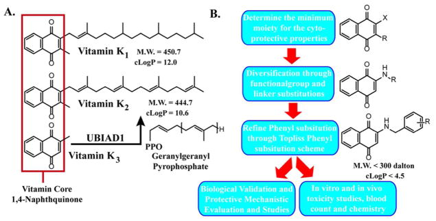
Historically known for its role in blood coagulation and bone formation, vitamin K (VK) has begun to emerge as an important nutrient for brain function. While VK involvement in the brain has not been fully explored, it is well-known that oxidative stress plays a critical role in neurodegenerative diseases. It was recently reported that VK protects neurons and oligodendrocytes from oxidative injury and rescues Drosophila from mitochondrial defects associated with Parkinson's disease. In this study, we take a chemical approach to define the optimal and minimum pharmacophore responsible for the neuroprotective effects of VK. In doing so, we have developed a series of potent VK analogues with favorable drug characteristics that provide full protection at nanomolar concentrations in a well-defined model of neuronal oxidative stress. Additionally, we have characterized key cellular responses and biomarkers consistent with the compounds' ability to rescue cells from oxidative stress induced cell death.
Figures
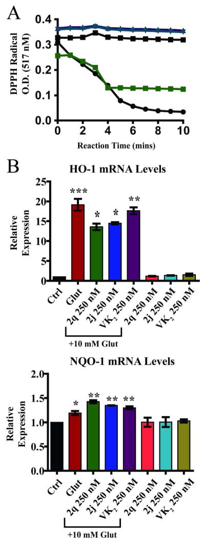
 ) and Trolox (●) used as controls. VK2 (■), 2j (
 ), and 2q (
 ) did not show direct antioxidant capacity. All compounds tested at 20 μM B. Expression of antioxidant response genes. Significant cellular antioxidant responses are elicited in HT22 cells after 8 hours of glutamate treatment with significant increase in HO-1 and NQO-1 gene expression. VK2, 2q, and 2j significantly decreased HO-1 expression but did not affect NQO-1 expression. One-way ANOVA with Bonferroni’s posttest was used to compare mean levels (n = 3), p<.01.
Similar articles
-
Apoptosis provoked by the oxidative stress inducer menadione (Vitamin K(3)) is mediated by the Fas/Fas ligand system.Clin Immunol. 1999 Oct;93(1):65-74. doi: 10.1006/clim.1999.4757. Clin Immunol. 1999. PMID: 10497012
-
Novel role of vitamin k in preventing oxidative injury to developing oligodendrocytes and neurons.J Neurosci. 2003 Jul 2;23(13):5816-26. doi: 10.1523/JNEUROSCI.23-13-05816.2003. J Neurosci. 2003. PMID: 12843286 Free PMC article.
-
Vitamin K protects against 7,12-dimethylbenz(A)anthracene induced hepatotoxicity in Wistar rats.Environ Toxicol. 2021 Mar;36(3):362-373. doi: 10.1002/tox.23042. Epub 2020 Oct 16. Environ Toxicol. 2021. PMID: 33063951
-
[Vitamin K metabolism. Menaquinone-4 (MK-4) formation from ingested VK analogues and its potent relation to bone function].Clin Calcium. 2007 Nov;17(11):1663-72. Clin Calcium. 2007. PMID: 17982185 Review. Japanese.
-
Vitamin K: New insights related to senescence and cancer metastasis.Biochim Biophys Acta Rev Cancer. 2024 Mar;1879(2):189057. doi: 10.1016/j.bbcan.2023.189057. Epub 2023 Dec 28. Biochim Biophys Acta Rev Cancer. 2024. PMID: 38158025 Review.
Cited by
-
Vitamin K2 in multiple sclerosis patients.Wien Klin Wochenschr. 2018 May;130(9-10):307-313. doi: 10.1007/s00508-018-1328-x. Epub 2018 Mar 2. Wien Klin Wochenschr. 2018. PMID: 29500722 Free PMC article.
-
Naphthoquinones as a Promising Class of Compounds for Facing the Challenge of Parkinson's Disease.Pharmaceuticals (Basel). 2023 Nov 8;16(11):1577. doi: 10.3390/ph16111577. Pharmaceuticals (Basel). 2023. PMID: 38004442 Free PMC article. Review.
-
Metal-Free Direct Amidation of Naphthoquinones Using Hydroxamic Acids as an Amide Source: Application in the Synthesis of an HDAC6 Inhibitor.Org Lett. 2016 Nov 4;18(21):5512-5515. doi: 10.1021/acs.orglett.6b02740. Epub 2016 Oct 19. Org Lett. 2016. PMID: 27759399 Free PMC article.
-
Novel Vitamin K analogs suppress seizures in zebrafish and mouse models of epilepsy.Neuroscience. 2014 Feb 14;259:142-54. doi: 10.1016/j.neuroscience.2013.11.040. Epub 2013 Dec 1. Neuroscience. 2014. PMID: 24291671 Free PMC article.
-
Role of SIRT1/PGC-1α in mitochondrial oxidative stress in autistic spectrum disorder.Neuropsychiatr Dis Treat. 2017 Jun 23;13:1633-1645. doi: 10.2147/NDT.S129081. eCollection 2017. Neuropsychiatr Dis Treat. 2017. PMID: 28694700 Free PMC article.
References
-
- Beckman KB, Ames BN. The free radical theory of aging matures. Physiol Rev. 1998;78:547–581. - PubMed
-
- Simonian NA, Coyle JT. Oxidative stress in neurodegenerative diseases. Annu Rev Pharmacol Toxicol. 1996;36:83–106. - PubMed
-
- Halliwell B. Role of free radicals in the neurodegenerative diseases: therapeutic implications for antioxidant treatment. Drugs Aging. 2001;18:685–716. - PubMed
-
- Riederer P, Sofic E, Rausch WD, Schmidt B, Reynolds GP, Jellinger K, Youdim MB. Transition metals, ferritin, glutathione, and ascorbic acid in parkinsonian brains. J Neurochem. 1989;52:515–520. - PubMed
-
- Sofic E, Lange KW, Jellinger K, Riederer P. Reduced and oxidized glutathione in the substantia nigra of patients with Parkinson’s disease. Neurosci Lett. 1992;142:128–130. - PubMed
Publication types
MeSH terms
Substances
Grants and funding
LinkOut - more resources
Full Text Sources
Other Literature Sources
Medical
