. 2013 Jan;4(1):80-93.
doi: 10.18632/oncotarget.793.

Genomic imbalance of HMMR/RHAMM regulates the sensitivity and response of malignant peripheral nerve sheath tumour cells to aurora kinase inhibition

Affiliations

Genomic imbalance of HMMR/RHAMM regulates the sensitivity and response of malignant peripheral nerve sheath tumour cells to aurora kinase inhibition

Pooja Mohan et al. Oncotarget. 2013 Jan.

Abstract

Malignant peripheral nerve sheath tumours (MPNST) are rare, hereditary cancers associated with neurofibromatosis type I. MPNSTs lack effective treatment options as they often resist chemotherapies and have high rates of disease recurrence. Aurora kinase A (AURKA) is an emerging target in cancer and an aurora kinase inhibitor (AKI), termed MLN8237, shows promise against MPNST cell lines in vitro and in vivo. Here, we test MLN8237 against two primary human MPNST grown in vivo as xenotransplants and find that treatment results in tumour cells exiting the cell cycle and undergoing endoreduplication, which cumulates in stabilized disease. Targeted therapies can often fail in the clinic due to insufficient knowledge about factors that determine tumour susceptibilities, so we turned to three MPNST cell-lines to further study and modulate the cellular responses to AKI. We find that the sensitivity of cell-lines with amplification of AURKA depends upon the activity of the kinase, which correlates with the expression of the regulatory gene products TPX2 and HMMR/RHAMM. Silencing of HMMR/RHAMM, but not TPX2, augments AURKA activity and sensitizes MPNST cells to AKI. Furthermore, we find that AURKA activity is critical to the propagation and self-renewal of sphere-enriched MPNST cancer stem-like cells. AKI treatment significantly reduces the formation of spheroids, attenuates the self-renewal of spheroid forming cells, and promotes their differentiation. Moreover, silencing of HMMR/RHAMM is sufficient to endow MPNST cells with an ability to form and maintain sphere culture. Collectively, our data indicate that AURKA is a rationale therapeutic target for MPNST and tumour cell responses to AKI, which include differentiation, are modulated by the abundance of HMMR/RHAMM.


Figures
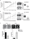
Figure 1
Figure 1. MLN8237 is effective against primary MPNSTs grown as xenotransplants in vivo
A. Sporadic MPNST tumours were orthotopically transplanted into NOD/SCID mice and allowed to grow to 2000 mm3 before treatment of 30mg/kg MLN8237 or vehicle was delivered daily. Treatment with MLN8237 resulted in stable disease after 2 weeks treatment, as determined by calliper measurements of tumour volumes, n=30 MLN8237 treated and n=14 for vehicle treated, *p<0.01, error bars = SD. Representative images of SP-MPNST tumours after 28 days of MLN8237 treatment or vehicle control are shown and treatment with the AKI resulted in significant reduction in tumour weight (quantitation on the right) (*p<0.01, Error bars = SD).B. NF1-MPNST tumours also show significantly stabilized tumour volumes and reduced tumour weight in the MLN8237 treated tumours compared to the vehicle controls. Vehicle treated mice, n=18, MLN8237 treated mice, n=21, error bars = SD. C. After 28 days of treatment, SP-MPNST tumour sections were stained for Ki-67 and DAPI. Ki-67 staining was significantly decreased in treated tumours suggesting that these cells have exited the cell cycle. Furthermore, there was a significant increase in multi-nucleate cells in treated tumours suggesting that cells are undergoing G2/M arrests and endoreduplication post treatment. *p<0.01, error bars = SD. Images are taken at 20x magnification.
Figure 2
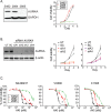
Figure 2. Inhibition of AURKA attenuates the growth of MPNST cell-lines in vitro
A. Cell lysates collected from S462, 2884 and 2885 cell lines show equivalent AURKA expression in S462 and 2884 cells lines. Furthermore, these cells lines undergo equivalent growth while 2885 cells experience slow proliferation. Seeding densities for S462 and 2884 were modified to enable equivalent proliferation. Cell viability was measured by MTT assays over a 4 day period. B. Immunoblot analysis of AURKA expression in lysates from untreated S462 cells and those treated with scrambled or AURKA targeted siRNA at 48 hours post transfection reveal specific reduction of AURKA. β-actin serves as a loading control. Cell viability is decreased in a dose dependent manner in cells treated with siRNA targeting AURKA relative to untreated cells and those cells treated with scrambled siRNA (right). Plotted siAurora values are obtained from four redundant siRNA targeting AURKA. Error bars = SEM, n=5 replicate experiments. C. Treatment of MPNST cells with three inhibitors to Aurora kinases, MLN8237, VX680 and C1368, reveals marked and dose-dependent reduction in cell viability as measured by MTT after 72 hours of treatment. Error bars = SEM, n=3 replicate experiments.
Figure 3
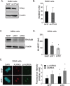
Figure 3. Gene dose alterations in TPX2 and RHAMM account for the differential AURKA activity in MPNST cell-lines
A. Augmented AURKA activity is detected in S462 cell lysates as measured by immunoblot detection of pS10-Histone H3B and pT288-AURKA. Apart from an increased level of RHAMM expression in 2884, levels of the unphosphorylated proteins are relatively constant. β-actin serves as a loading control. B. Comparative genomic hybridization of genomic DNA from S462, 2884 and 2885 MPNST cell lines identify copy number gains of 20q in S462 and 2884 cells, and a gain of 5q33.2-qter in 2884 cells. C. Quantitative PCR confirms increased gene dosage of TPX2, but decreased HMMR, in S462 relative to 2884 cells. Error bars =SD, n=3. D. Gene dosage translates to different levels of mRNA for TPX2 and RHAMM as detected by qRT-PCR in S462 relative to 2884 cells. Error bars=SD, n=3. E. Representative blots for protein expression of AURKA, TPX2 and RHAMM as detected in cell lysates from S462 and 2884 cell-lines by IR labelled antibodies. Consistent with the genomic and message levels, S462 cells contain a marked increase in TPX2 expression and a reduced expression of RHAMM relative to 2884 cells.
Figure 4
Figure 4. RHAMM depletion increases AURKA activity and sensitivity to AKI in 2884 cells
A. TPX2 was stably knocked down in S462 cells using shRNA mediated silencing of either TPX2 or a control non-hairpin (NHP) construct. β-actin served as a loading control. B. shTPX2 S462 cells experienced no significant differences in IC-50 to MLN8237 compared to NHP cells, error bars = SD, n=3. C. RHAMM was knocked down in 2884 cells with a similar shRNA expression system. Two redundant shRNAs were used, shR1 and shR2. β-actin served as a loading control. D. shR1 and shR2 cells experienced a 2 fold decrease in IC-50 in response to MLN8237 treatment compared to NHP cells, *p value<0.05, error bars = SD, n=3. E. Representative images of NHP and shR1 cells at metaphase stained for AURKA and pAURKA-T288. Scale bars = 5μm. Quantitation of fluorescent intensity of spindle poles was done using FV10-ASW software, *p value <0.05, error bars = SD.
Figure 5
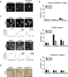
Figure 5. AKI treatment results in growth arrest, apoptosis, polyploidy and cellular senescence in MPNST cell lines
A. Treatment of S462 and 2884 cells with AKI for 48 hours resulted in low levels of apoptosis as detected by Annexin V staining by high content cell screening. Representative images are shown with non-viable, membrane permeable cells counter stained with propidium iodide (PI). B. Quantitation of cellular responses to AKI or vehicle treatments, *p value <0.05, error bars = SD, n=3 for apoptosis measurements, n=3 for polyploidy and n=4 for senescence quantitation. C. S462 and 2884 cells were exposed to AKIs, or vehicle control, and changes in the nuclear content of treated cells was detected by PI staining and FACS analysis. S462 cells exposed to AKIs display larger G2/M and 8N fractions, indicative of an endoreduplication phenotype. Representative images of cells stained with DAPI are shown above the FACS profiles to illustrate the multi-nucleate phenotypes. D. Treatment of S462 cells with AKIs at IC-50 concentrations for 72 hours induces senescence activated β-gal expression in comparison to DMSO treated cells. Representative images were taken on an Axiovert 40 microscope (Zeiss).
Figure 6
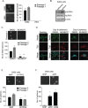
Figure 6. Inhibition of AURKA by MLN8237 limits self-renewal and induces neuronal differentiation of MPNST tumour initiating cells in vitro
A. S462 and 2884 cells were cultured in anchorage independent conditions in neurocult media and sphere formation was quantitated after 6 days (passage 0). Spheres were dissociated and cultured for a further 6 days and sphere formation was measured again (passage 1). S462 cells form spheres whereas, 2884 cells do not. Representative images on the left, scale bars = 50μm, quantitation on the right, error bars = SD, n=3. B. In comparison to adherent S462 cell lysates, sphere-enriched S462 lysates (sphere) contain higher levels of auto-activated pAURKA (Thr288) indicating increased AURKA activity in sphere versus adherent S462 cells. β-actin serves as a loading control. C. In comparison to DMSO (vehicle) treated controls, prolonged treatment of S462 with 100 nM MLN8237 inhibits the propagation (passage 0) and self-renewal (passage 1) of S462 cells cultured as spheres. Quantitation is shown below representative images. Scale bars equal 50 μm. Error bars = SD, n=3, *p<0.05, **p<0.001. D. Dissociated sphere-enriched cells at passage 4 were fixed and stained for DAPI, nestin, and Tuj1 at day 0 and following 9 days of treatment with MLN8237 (50nM), or DMSO. Cells grew in both adherence and sphere phenotypes. While untreated cells maintain expression of nestin through the nine days of culture, MLN8237 treated spheres lost nestin expression and are positive for the neuronal marker, Tuj1 instead. Scale bars equal 50μm. E. shTPX2 and NHP S462 cells were grown in neurocult media. While there were no significant differences in sphere formation in passage 0, TPX2 knockdown is effecting the self-renewal of these spheres in passage 1, scale bars = 50μm, *p-value< 0.05, error bars = SD, n=3. F. shR1 and shR2 cells form significantly more spheres in neurocult media than NHP 2884 cells, indicating a role for RHAMM in sphere formation by modulating AURKA activity. Scale bars = 50μm, *p-value < 0.05, error bars = SD, n=3.

Similar articles

Cited by

References

    1. Jett K, Friedman JM. Clinical and genetic aspects of neurofibromatosis 1. Genet Med. 2010;12(1):1–11. - PubMed
    1. Brems H, Beert E, de Ravel T, Legius E. Mechanisms in the pathogenesis of malignant tumours in neurofibromatosis type 1. Lancet Oncol. 2009;10(5):508–515. - PubMed
    1. Woodruff JM. Pathology of tumors of the peripheral nerve sheath in type 1 neurofibromatosis. Am J Med Genet. 1999;89(1):23–30. - PubMed
    1. Hagel C, Zils U, Peiper M, Kluwe L, Gotthard S, Friedrich RE, Zurakowski D, von Deimling A, Mautner VF. Histopathology and clinical outcome of NF1-associated vs. sporadic malignant peripheral nerve sheath tumors. J Neurooncol. 2007;82(2):187–192. - PubMed
    1. Patel AV, Eaves D, Jessen WJ, Rizvi TA, Ecsedy JA, Qian MG, Aronow B, Perentesis J, Serra E, Cripe T, Miller SC, Ratner N. Ras-Driven Transcriptome Analysis Identifies Aurora Kinase A as a Potential Malignant Peripheral Nerve Sheath Tumor Therapeutic Target. Clin Cancer Res. 2012 - PMC - PubMed

Publication types

MeSH terms