. 2013 Mar 15;304(6):G615-22.
doi: 10.1152/ajpgi.00445.2012. Epub 2013 Jan 17.

Keratinocyte growth factor-2 stimulates P-glycoprotein expression and function in intestinal epithelial cells

Affiliations

Keratinocyte growth factor-2 stimulates P-glycoprotein expression and function in intestinal epithelial cells

Seema Saksena et al. Am J Physiol Gastrointest Liver Physiol. .

Abstract

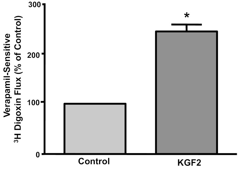
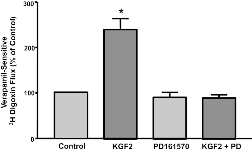
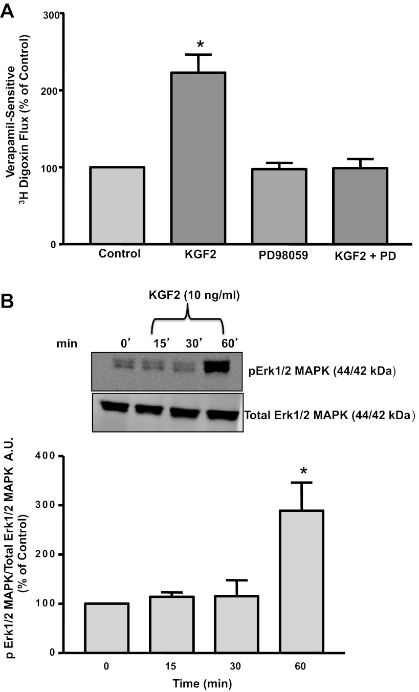
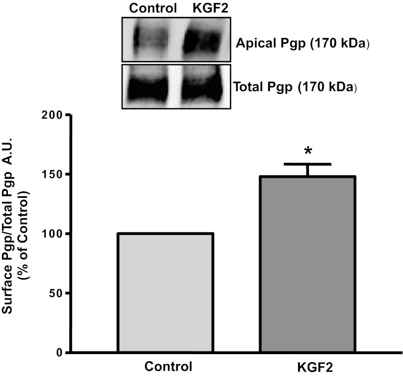
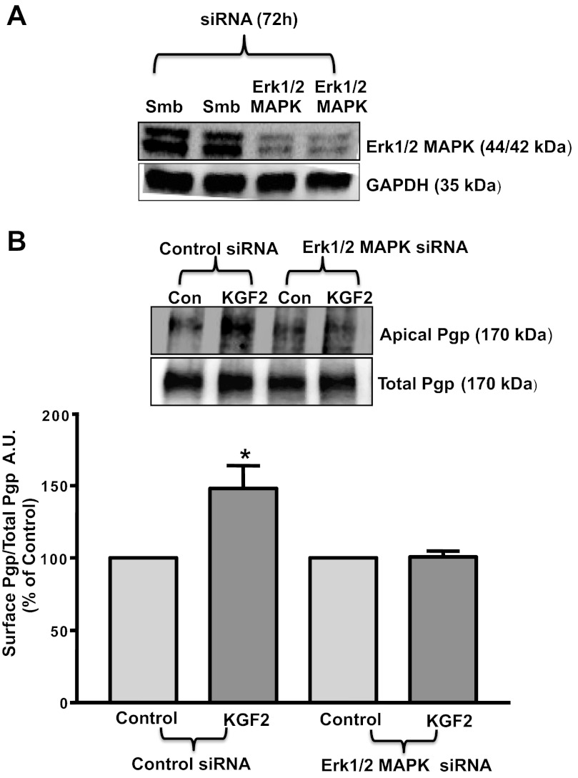
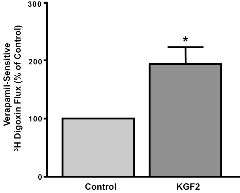
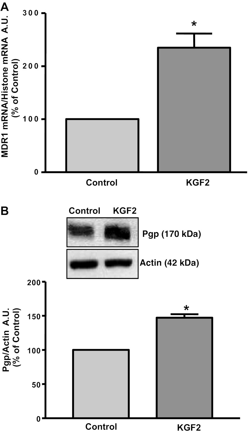
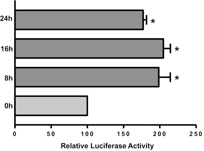
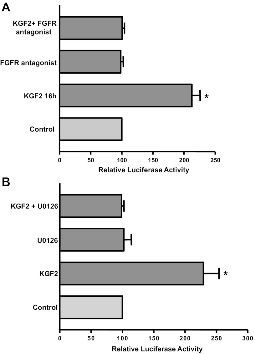
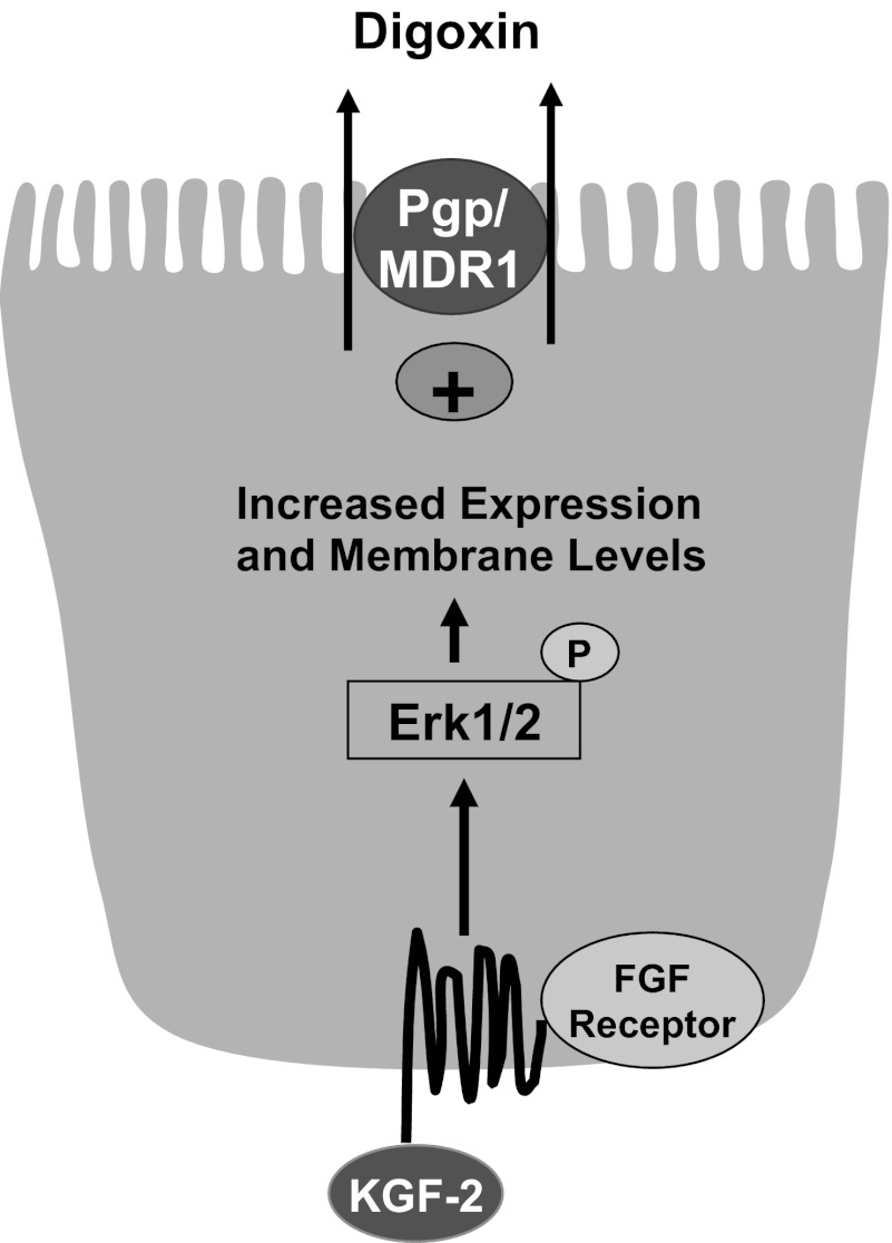
Intestinal P-glycoprotein (Pgp/multidrug resistance 1), encoded by the ATP-binding cassette B1 gene, is primarily involved in the transepithelial efflux of toxic metabolites and xenobiotics from the mucosa into the gut lumen. Reduced Pgp function and expression has been shown to be associated with intestinal inflammatory disorders. Keratinocyte growth factor-2 (KGF2) has emerged as a potential target for modulation of intestinal inflammation and maintenance of gut mucosal integrity. Whether KGF2 directly regulates Pgp in the human intestine is not known. Therefore, the present studies were undertaken to determine the modulation of Pgp by KGF2 using Caco-2 cells. Short-term treatment of Caco-2 cells with KGF2 (10 ng/ml, 1 h) increased Pgp activity (~2-fold, P < 0.05) as measured by verapamil-sensitive [(3)H]digoxin flux. This increase in Pgp function was associated with an increase in surface Pgp levels. The specific fibroblast growth factor receptor (FGFR) antagonist PD-161570 blocked the KGF2-mediated increase in Pgp activity. Inhibition of the mitogen-activated protein kinase (MAPK) pathway by PD-98059 attenuated the stimulatory effects of KGF2 on Pgp activity. Small-interfering RNA knockdown of Erk1/2 MAPK blocked the increase in surface Pgp levels by KGF2. Long-term treatment with KGF2 (10 ng/ml, 24 h) also significantly increased PgP activity, mRNA, protein expression, and promoter activity. The long-term effects of KGF2 on Pgp promoter activity were also blocked by the FGFR antagonist and mediated by the Erk1/2 MAPK pathway. In conclusion, our findings define the posttranslational and transcriptional mechanisms underlying stimulation of Pgp function and expression by KGF2 that may contribute to the beneficial effects of KGF2 in intestinal inflammatory disorders.


Figures

Fig. 1.
Fig. 1.
Short-term effect of keratinocyte growth factor-2 (KGF2) on P-glycoprotein (Pgp) activity in Caco-2 cells. Overnight serum-starved postconfluent Caco-2 cells were treated with KGF2 (10 ng/ml) in serum-free cell culture medium supplemented with 0.2% BSA for 60 min. [3H]digoxin flux (1 μM) was measured as described in materials and methods. Results are expressed as %control and represent means ± SE of 6 separate experiments performed in triplicate. *P < 0.05 compared with untreated control.
Fig. 2.
Fig. 2.
Effects of KGF2 on Pgp activity in Caco-2 cells are receptor mediated. Postconfluent Caco-2 cells were pretreated with specific fibroblast growth factor (FGF) receptor antagonist PD-161570 (10 μM) for 60 min in the serum-free cell culture medium and then coincubated with KGF2 (10 ng/ml) in serum-free cell culture medium supplemented with 0.2% BSA for 60 min. [3H]digoxin flux (1 μM) was measured as described in materials and methods. Results are expressed as %control and represent means ± SE of 4 separate experiments performed in triplicate. *P < 0.05 compared with untreated control.
Fig. 3.
Fig. 3.
A: effect of Erk1/2 mitogen-activated protein kinase (MAPK) inhibitor on KGF2-induced stimulation of Pgp activity in Caco-2 cells. Postconfluent Caco-2 cells were pretreated with the specific inhibitor of Erk1/2 MAPK PD-98059 (30 μM) for 60 min in the serum-free cell culture medium and then coincubated with KGF2 (10 ng/ml) in serum-free cell culture medium supplemented with 0.2% BSA for 60 min. [3H]digoxin flux (1 μM) was measured as described in materials and methods. Results are expressed as %control and represent means ± SE of 5 separate experiments performed in triplicate. *P < 0.05 compared with untreated control. B: KGF2 induces Erk1/2 MAPK phosphorylation in Caco-2 cells. Caco-2 cells were incubated with KGF2 (10 ng/ml) in serum-free cell culture medium supplemented with 0.2% BSA for different time intervals ranging from 15 to 60 min. After the cells were washed with 1× PBS, extracted proteins (75 μg) were subjected to Western blot analysis on 12% SDS-polyacrylamide gel using phospho-specific Erk1/2 MAPK antibody (pErk1/2 MAPK). The blots were stripped and reprobed with the ERK1/2 MAPK antibody (total Erk1/2 MAPK) to indicate equal loading of protein in each lane. A representative blot of 3 different experiments is shown. The data were quantified by densitometric analysis and expressed as arbitrary units and represent means ± SE of 3 separate experiments. *P < 0.05 compared with untreated control (0 min).
Fig. 4.
Fig. 4.
KGF2 increases Pgp surface expression. Overnight serum-starved postconfluent Caco-2 cells were treated with KGF2 (10 ng/ml) in serum-free cell culture medium supplemented with 0.2% BSA for 60 min and subjected to biotinylation at 4°C using sulfo-NHS-SS-biotin. After solubilization, biotinylated proteins were extracted with streptavidin-agarose from equal amounts of total cellular protein. Surface and total cellular fractions were run on 7% SDS-polyacrylamide gel electrophoresis followed by transfer to nitrocellulose membrane. The blot was immunostained with a mouse anti-Pgp/multidrug resistance 1 [MDR1 (Pgp)]. A representative blot of 3 different experiments is shown. The data were quantified by densitometric analysis and expressed as arbitrary units and represent means ± SE of 3 separate experiments.
Fig. 5.
Fig. 5.
Erk1/2 MAPK knockdown abrogates KGF2-induced increase in Pgp surface expression. A: Erk1/2 MAPK small-interfering RNA (siRNA) causes knockdown of Erk1/2 MAPK expression in Caco-2 cells. Scrambled (Smb) or Erk1/2 MAPK siRNA-transfected Caco-2 cell lysates (75 μg) were subjected to 12% SDS-PAGE followed by transfer to nitrocellulose membrane. The blot was probed with anti-Erk1/2 MAPK antibody (Erk1/2 MAPK) or anti-GAPDH (GAPDH) antibody. B: cell surface biotinylation in siRNA-transfected Caco-2 cells. Overnight serum-starved cells transfected with scrambled siRNA or with Erk1/2 MAPK siRNA were treated with KGF2 (10 ng/ml) in serum-free cell culture medium supplemented with 0.2% BSA for 60 min and subjected to biotinylation at 4°C using sulfo-NHS-SS-biotin. After solubilization, biotinylated proteins were extracted with streptavidin-agarose from equal amounts of total cellular protein. Surface and total cellular fractions were run on 7% SDS-polyacrylamide gel electrophoresis followed by transfer to nitrocellulose membrane. The blot was immunostained with a mouse anti-Pgp/MDR1 (Pgp). A representative blot of 3 different experiments is shown. The data were quantified by densitometric analysis and expressed as arbitrary units and represent means ± SE of 3 separate experiments.
Fig. 6.
Fig. 6.
Long-term effect of KGF2 on Pgp activity in Caco-2 cells. Overnight serum-starved postconfluent Caco-2 cells were treated with KGF2 (10 ng/ml) in serum-free cell culture medium supplemented with 0.2% BSA for 24 h. [3H]digoxin flux (1 μM) was measured as described in materials and methods. Results are expressed as %control and represent means ± SE of 5 separate experiments performed in triplicate. *P < 0.05 compared with untreated control.
Fig. 7.
Fig. 7.
KGF2 induces Pgp mRNA and protein expression in Caco-2 cells. Overnight serum-starved postconfluent Caco-2 cells were treated with KGF2 (10 ng/ml) in serum-free cell culture medium supplemented with 0.2% BSA for 24 h. A: total RNA was then extracted from the cells, and 100 ng were amplified with MDR1 or histone gene-specific primers using 1-step RT-PCR mix containing SYBR Green fluorescence dye for real-time PCR quantitation. The relative abundance of MDR1 mRNA from control and KGF2-treated Caco-2 cells was normalized to histone mRNA (internal control). The data were quantified by densitometric analysis and expressed as %control in arbitrary units. Results represent means ± SE of 3 independent experiments performed in triplicate. *P < 0.05 compared with untreated control. B: control and KGF2-treated cell lysates (75 μg) were subjected to 7% SDS-PAGE followed by transfer to nitrocellulose membrane. The blot was probed with anti-Pgp/MDR1 (Pgp) or anti-β-actin (Actin) antibody. A representative blot of 3 different experiments is shown. The data were quantified by densitometric analysis and expressed as %control in arbitrary units. Results represent means ± SE of 3 independent experiments performed in triplicate. *P < 0.05 compared with untreated control.
Fig. 8.
Fig. 8.
Effect of KGF2 on Pgp promoter activity in Caco-2 cells. Caco-2 cells were transiently transfected with MDR1 luciferase promoter construct (p-1,073/+703) along with pCMVβ vector. Transfected cells were treated with KGF2 (10 ng/ml) for different periods of time in media containing 0.2% BSA and 1% FBS after 24 h. Posttransfection (48 h), the promoter activity was measured by luciferase assay. Values were normalized to β-galactosidase activity to correct for transfection efficiency. Results represent means ± SE of 3 separate experiments and are expressed as %control comparing transfected cells treated with KGF2 with untreated cells (control). *P < 0.05 compared with control.
Fig. 9.
Fig. 9.
Effect of FGF receptor antagonist or Erk1/2 MAPK inhibitor on KGF2-induced stimulation of Pgp promoter activity in Caco-2 cells. Caco-2 cells were transiently transfected with MDR1 luciferase promoter construct (p-1,073/+703) along with pCMVβ vector. After 24 h, transiently transfected cells were pretreated with the specific antagonist of FGF receptor PD-161570 (10 μM) (A) or the Erk1/2 MAPK inhibitor U-0126 (10 μM) (B) for 60 min in media containing 0.2% BSA and 1% FBS and then coincubated with KGF2 (10 ng/ml) in media containing 1% FBS for 24 h. Posttransfection (48 h) the promoter activity was measured by luciferase assay. Values were normalized to β-galactosidase activity to correct for transfection efficiency. Results represent means ± SE of 3 separate experiments and are expressed as %control comparing transfected cells treated with KGF2 with untreated cells (control). *P < 0.05 compared with control.
Fig. 10.
Fig. 10.
Schematic of the proposed model of KGF2-mediated effects on Pgp.

Similar articles

Cited by

References

    1. Annese V, Valvano MR, Palmieri O, Latiano A, Bossa F, Andriulli A. Multidrug resistance 1 gene in inflammatory bowel disease: a meta-analysis. World J Gastroenterol 12: 3636–3644, 2006 - PMC - PubMed
    1. Asaki T, Konishi M, Miyake A, Kato S, Tomizawa M, Itoh N. Roles of fibroblast growth factor 10 (Fgf10) in adipogenesis in vivo. Mol Cell Endocrinol 218: 119–128, 2004 - PubMed
    1. Avissar NE, Sax HC, Toia L. In human entrocytes, GLN transport and ASCT2 surface expression induced by short-term EGF are MAPK, PI3K, and Rho-dependent. Dig Dis Sci 53: 2113–2125, 2008 - PubMed
    1. Blaisdell CJ, Pellettieri JP, Loughlin CE, Chu S, Zeitlin PL. Keratinocyte growth factor stimulates CLC-2 expression in primary fetal rat distal lung epithelial cells. Am J Respir Cell Mol Biol 20: 842–847, 1999 - PubMed
    1. Blokzijl H, Vander Borght S, Bok LI, Libbrecht L, Geuken M, van den Heuvel FA, Dijkstra G, Roskams TA, Moshage H, Jansen PL, Faber KN. Decreased P-glycoprotein (P-gp/MDR1) expression in inflamed human intestinal epithelium is independent of PXR protein levels. Inflamm Bowel Dis 13: 710–720, 2007 - PubMed

Publication types

MeSH terms

LinkOut - more resources