Randomized Controlled Trial
. 2013 Jul:5:40-50.
doi: 10.1016/j.dcn.2012.12.001. Epub 2012 Dec 27.

Dimensional change card sort performance associated with age-related differences in functional connectivity of lateral prefrontal cortex

Affiliations
Randomized Controlled Trial

Dimensional change card sort performance associated with age-related differences in functional connectivity of lateral prefrontal cortex

Fredrick Ezekiel et al. Dev Cogn Neurosci. 2013 Jul.

Abstract

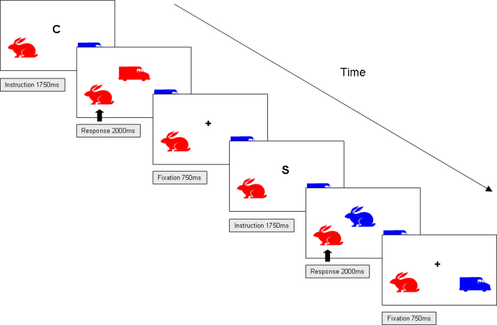
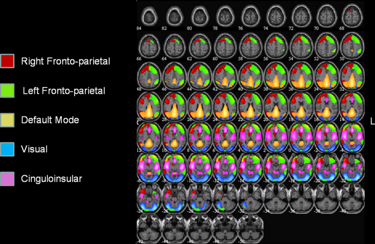
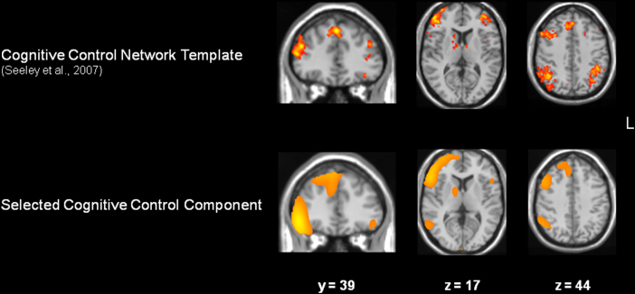
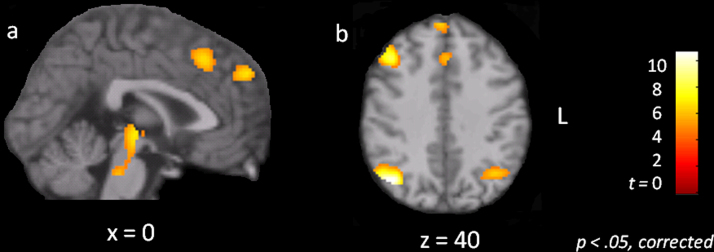
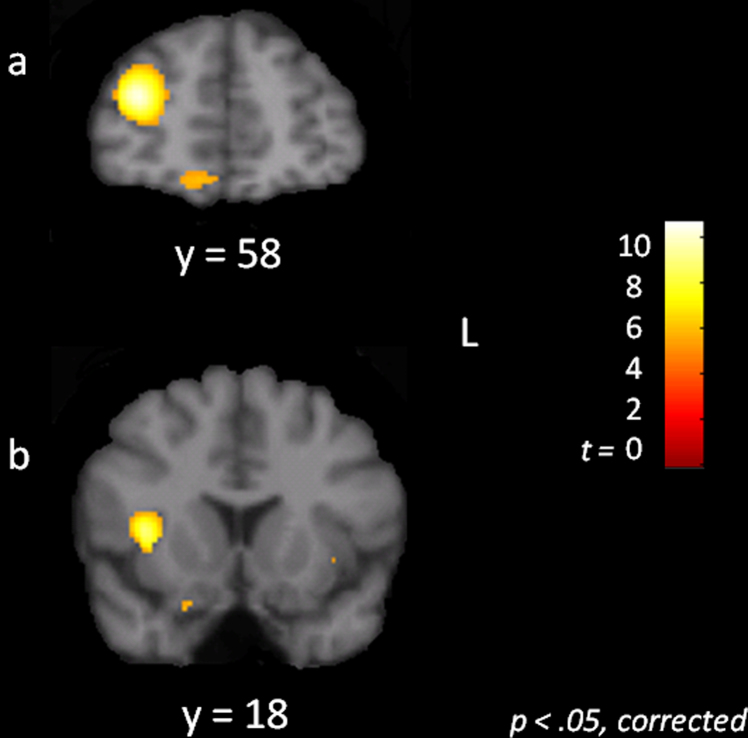
The Dimensional Change Card Sort (DCCS) is a standard procedure for assessing executive functioning early in development. In the task, participants switch from sorting cards one way (e.g., by color) to sorting them a different way (e.g., by shape). Traditional accounts associate age-related changes in DCCS performance with circumscribed changes in lateral prefrontal cortex (lPFC) functioning, but evidence of age-related differences in the modulation of lPFC activity by switching is mixed. The current study therefore tested for possible age-related differences in functional connectivity of lPFC with regions that comprise a larger cognitive control network. Functional magnetic resonance imaging (fMRI) data collected from children and adults performing the DCCS were analyzed by means of independent components analysis (ICA). The analysis revealed several important age-related differences in functional connectivity of lPFC. In particular, lPFC was more strongly connected with the anterior cingulate, inferior parietal cortex, and the ventral tegmental area in adults than in children. Theoretical implications are discussed.


Figures

Fig. 1
Fig. 1
An illustration of two representative trials from the modified Dimensional Change Card Sort task used in the present study. Trials began with an instruction cue indicating the rule on that trial, followed by the presentation of a stimulus to which participants responded, followed by a fixation point. On switch trials, the rule was different than on the previous trial; on repeat trials, the rule was the same as on the previous trial. Individual trials were administered in the form of a block design.
Fig. 2
Fig. 2
A composite view of five group components yielded by the ICA decomposition. Group components were converted to Z-scores and then mapped for the purpose of visualization at a threshold of Z > 1.
Fig. 3
Fig. 3
Cognitive control network template (from Seeley et al., 2007) and the selected right fronto-parietal component. Spatial correlation confirmed the strong correspondence of the two maps (p < .05), which was especially evident in lateral prefrontal, anterior cingulate/medial prefrontal cortex, dorsal striatum, and parietal cortex.
Fig. 4
Fig. 4
Statistical parametric maps showing voxels more strongly connected to the cognitive control network in adults as compared to children, p < .05, corrected. (a) Sagittal slice showing group differences in the anterior cingulate cortex, the medial superior frontal gyrus, pulvinar, and ventral tegmental area. (b) Axial slice showing group differences in right dorsolateral prefrontal cortex, anterior cingulate cortex, medial superior frontal gyrus, and inferior parietal cortex bilaterally.
Fig. 5
Fig. 5
Statistical parametric maps showing voxels more strongly connected to the cognitive control network in children as compared to adults, p < .05, corrected. There was no evidence of stronger lPFC connectivity with the cognitive control network in children compared to adults. (a) Coronal slice showing group differences in the anterior extent of the inferior and middle frontal gyri/frontal pole. (b) Coronal slice showing group differences in the right anterior insula.

References

    1. Alexander G.E., DeLong M.R., Strick P.L. Parallel organization of functionally segregated circuits linking basal ganglia and cortex. Annual Review of Neuroscience. 1986;9:357–381. - PubMed
    1. Allen E.A., Erhardt E.B., Damaraju E., Gruner W., Segall J.M., Silva R.F. A baseline for the multivariate comparison of resting-state networks. Frontiers in Systems Neuroscience. 2011;5:2. - PMC - PubMed
    1. Allen E.A., Erhardt E.B., Wei Y., Eichele T., Calhoun V.D. Capturing inter-subject variability with group independent component analysis of fMRI data: a simulation study. Neuroimage. 2012;59(4):4141–4159. - PMC - PubMed
    1. Barber A.D., Carter C.S. Cognitive control involved in overcoming prepotent response tendencies and switching between tasks. Cerebral Cortex. 2005;15(7):899–912. - PubMed
    1. Barberi E.A., Gati J.S., Rutt B.K., Menon R.S. A transmit-only/receive-only (TORO) RF system for high-field MRI/MRS applications. Magnetic Resonance in Medicine. 2000;43(2):284–289. - PubMed

Publication types

LinkOut - more resources