

Earlier detection of breast cancer with ultrasound molecular imaging in a transgenic mouse model
- PMID: 23328585
- PMCID: PMC3602408
- DOI: 10.1158/0008-5472.CAN-12-3391
Earlier detection of breast cancer with ultrasound molecular imaging in a transgenic mouse model
Abstract
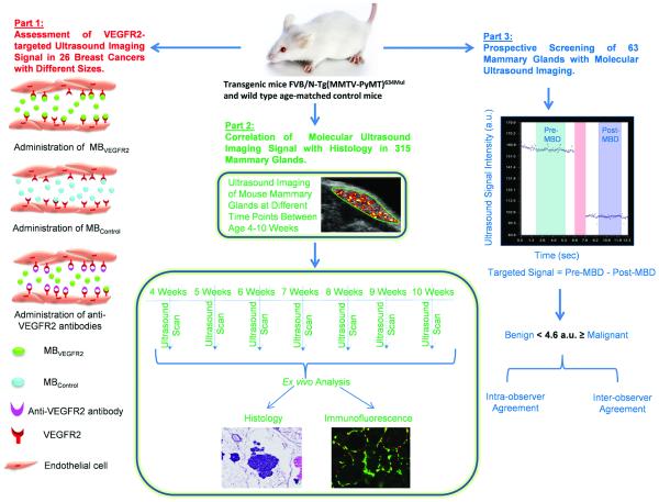
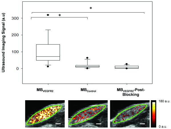
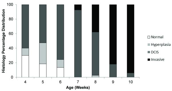
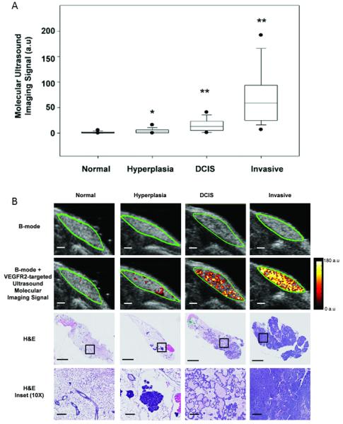
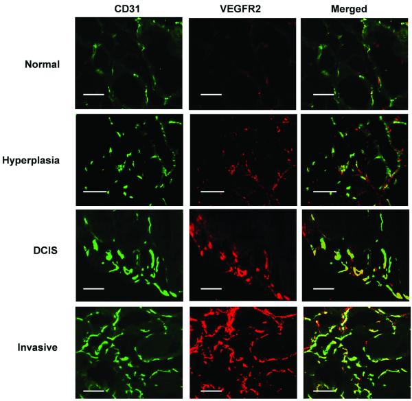
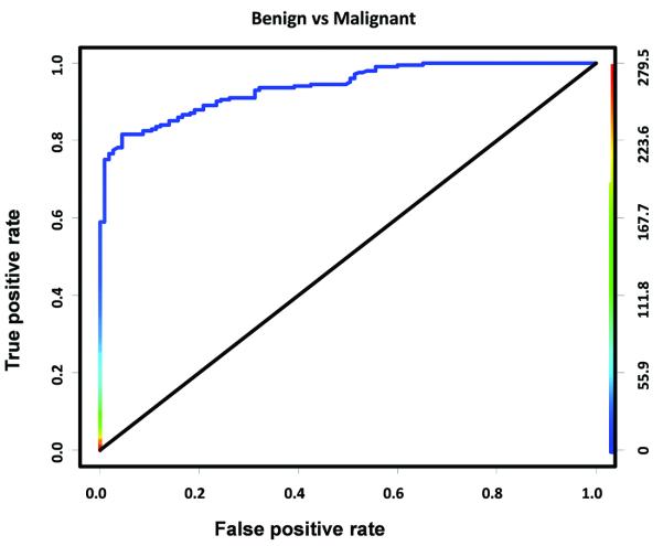
While there is an increasing role of ultrasound for breast cancer screening in patients with dense breast, conventional anatomical ultrasound lacks sensitivity and specificity for early breast cancer detection. In this study, we assessed the potential of ultrasound molecular imaging using clinically translatable vascular endothelial growth factor receptor type 2 (VEGFR2)-targeted microbubbles (MB(VEGFR2)) to improve the diagnostic accuracy of ultrasound in earlier detection of breast cancer and ductal carcinoma in situ (DCIS) in a transgenic mouse model [FVB/N-Tg(MMTV-PyMT)634Mul]. In vivo binding specificity studies (n = 26 tumors) showed that ultrasound imaging signal was significantly higher (P < 0.001) using MB(VEGFR2) than nontargeted microbubbles and imaging signal significantly decreased (P < 0.001) by blocking antibodies. Ultrasound molecular imaging signal significantly increased (P < 0.001) when breast tissue (n = 315 glands) progressed from normal [1.65 ± 0.17 arbitrary units (a.u.)] to hyperplasia (4.21 ± 1.16), DCIS (15.95 ± 1.31), and invasive cancer (78.1 ± 6.31) and highly correlated with ex vivo VEGFR2 expression [R(2) = 0.84; 95% confidence interval (CI), 0.72-0.91; P < 0.001]. At an imaging signal threshold of 4.6 a.u., ultrasound molecular imaging differentiated benign from malignant entities with a sensitivity of 84% (95% CI, 78-88) and specificity of 89% (95% CI, 81-94). In a prospective screening trail (n = 63 glands), diagnostic performance of detecting DCIS and breast cancer was assessed and two independent readers correctly diagnosed malignant disease in more than 95% of cases and highly agreed between each other [intraclass correlation coefficient (ICC) = 0.98; 95% CI, 97-99]. These results suggest that VEGFR2-targeted ultrasound molecular imaging allows highly accurate detection of DCIS and breast cancer in transgenic mice and may be a promising approach for early breast cancer detection in women.
Figures
References
-
- Siegel R, Naishadham D, Jemal A. Cancer statistics, 2012. CA Cancer J Clin. 2012;62:10–29. - PubMed
-
- Humphrey LL, Helfand M, Chan BK, Woolf SH. Breast cancer screening: a summary of the evidence for the U.S. Preventive Services Task Force. Ann Intern Med. 2002;137:347–60. - PubMed
-
- Stomper PC, D’Souza DJ, DiNitto PA, Arredondo MA. Analysis of parenchymal density on mammograms in 1353 women 25-79 years old. AJR Am J Roentgenol. 1996;167:1261–5. - PubMed
-
- Kolb TM, Lichy J, Newhouse JH. Comparison of the performance of screening mammography, physical examination, and breast US and evaluation of factors that influence them: an analysis of 27,825 patient evaluations. Radiology. 2002;225:165–75. - PubMed
-
- Mandelson MT, Oestreicher N, Porter PL, White D, Finder CA, Taplin SH, et al. Breast density as a predictor of mammographic detection: comparison of interval- and screen-detected cancers. J Natl Cancer Inst. 2000;92:1081–7. - PubMed
Publication types
MeSH terms
Substances
Grants and funding
LinkOut - more resources
Full Text Sources
Other Literature Sources
Medical
Molecular Biology Databases
Miscellaneous
