. 2013 Jan 17;4(1):e462.
doi: 10.1038/cddis.2012.214.

Mast cells rescue implantation defects caused by c-kit deficiency

Affiliations

Mast cells rescue implantation defects caused by c-kit deficiency

K Woidacki et al. Cell Death Dis. .

Abstract

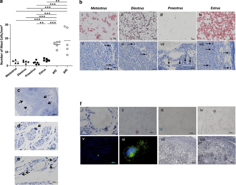
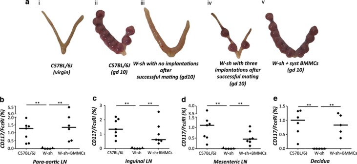
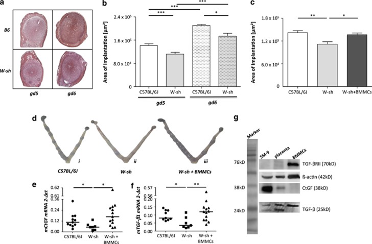
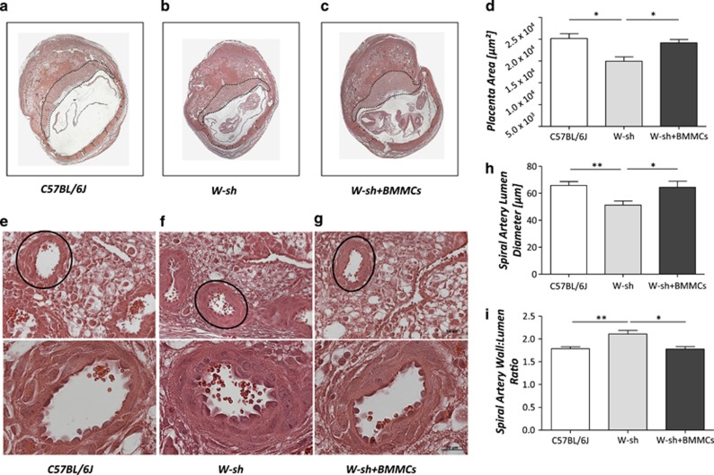
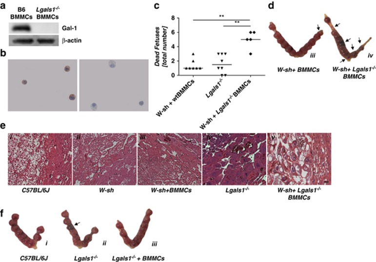
Various physiologically relevant processes are regulated by the interaction of the receptor tyrosine kinase (c-Kit) and its ligand stem cell factor (SCF), with SCF known to be the most important growth factor for mast cells (MCs). In spite of their traditional role in allergic disorders and innate immunity, MCs have lately emerged as versatile modulators of a variety of physiologic and pathologic processes. Here we show that MCs are critical for pregnancy success. Uterine MCs presented a unique phenotype, accumulated during receptivity and expanded upon pregnancy establishment. Kit(W-sh/W-sh) mice, whose MC deficiency is based on restricted c-Kit gene expression, exhibited severely impaired implantation, which could be completely rescued by systemic or local transfer of wild-type bone marrow-derived MCs. Transferred wild-type MCs favored normal implantation, induced optimal spiral artery remodeling and promoted the expression of MC proteases, transforming growth factor-β and connective tissue growth factor. MCs contributed to trophoblast survival, placentation and fetal growth through secretion of the glycan-binding protein galectin-1. Our data unveil unrecognized roles for MCs at the fetomaternal interface with critical implications in reproductive medicine.


Figures

Figure 1
Figure 1
The numbers of uterine MCs increase during the fertile period of the estrus cycle and remain high during early gestation. Female virgin C57BL/6J females were checked for their phase of the estrous cycle. (bi–iv) Characteristic pictures from hematoxylin/eosin (H/E)-stained vaginal lavage samples of each phase are shown. (bv–viii) The number of MCs in uterine paraffin sections (5 μm) of female virgin (n= 4–7), 2-day and 5-day pregnant BALB/c-mated C57BL/6J females (gd2 and gd5, n=5 each) was quantified in 1 mm2 (magnification × 200) areas by visualizing them with Toluidine blue O staining (0.1%). The mean MC number from both the right and left uterine horn was calculated and included in (a). At day 5 of pregnancy, MCs were located in between the implantation sites (c), whereas at day 10 of pregnancy, MCs were detected in the decidua, the maternal part of the fetomaternal interface, of BALB/c-mated C57BL/6J females (d) and are particularly localized close to blood vessels (e; magnification × 1000). (f) Uterine MCs that are positive for Toluidine blue (i), Safranin (ii), Alcian blue (iii) and double positive for Safranin and Alcian blue (iv). In the lower panel, uterine MCs are shown that are positive for CD117 (v and vi) or Mcpt-8 (vii and viii). i–iv and vi: magnification × 1000, v and vii: × 100 magnification and viii: × 400 magnification. Arrows indicate uterine MCs. Data are expressed as single dots with medians and were analyzed by using the Mann–Whitney U-test (*P<0.05; **P<0.01; ***P<0.001)
Figure 2
Figure 2
Systemic transfer of BMMCs reverses the low number of implantations of MC-deficient mice. KitW-sh/W-sh females (W-sh) were reconstituted systemically with BMMCs 12 weeks before being mated with BALB/c males. Implantation success was documented on day 10 of pregnancy. BALB/c-mated C57BL/6J females and KitW-sh/W-sh females served as controls. In (a) representative pictures are shown from uteri of (i) a virgin C57BL/6J female, (ii) a normal pregnant C57BL/6J, (iii) a non-pregnant KitW-sh/W-sh female with an inflamed and thickened uterus after successful mating, (iv) a pregnant KitW-sh/W-sh female presenting only three implantations and (v) a pregnant KitW-sh/W-sh female that was systemically reconstituted with BMMCs from wild-type mice showing 11 implantations. (bd) Percentage of CD117/FcɛRIα+ cells in para-aortic (b), inguinal (c) and mesenteric (d) lymph nodes from C57BL/6J (n=7); KitW-sh/W-sh (n=5) or KitW-sh/W-sh+BMMCs (n=6–7) animals. (e) Percentage of CD117/FcɛRIα+ cells in the decidua from C57BL/6J (n=6), KitW-sh/W-sh (n=5) and KitW-sh/W-sh+BMMCs (n=5) animals. Data are expressed as medians. *P<0.05, **P<0.005, ***P<0.001 as analyzed by Kruskall–Wallis test followed by Mann–Whitney Utest between two particular groups
Figure 3
Figure 3
Implantation areas of KitW-sh/W-sh (W-sh) females are significantly smaller than those of wild-type mice and this can be reversed by transfer of BMMCs; Normalization of the TGF-β/CtGF axis. (a) Representative images of implantations as recorded at gestation days (gd) 5 and 6 in wild-type C57/BL6 and KitW-sh/W-sh mice after Chicago Blue dye injection. The quantification of the area size (μm2) was done by AxioVision4 (Zeiss) and is shown in (b) the number of mice per group was as follows: C57/BL6 day 5: n=9 and day 6: n=4, KitW-sh/W-sh day 5: n=6 and day 6 n=4. (c and d) representative uteri (c) and the measured implantation areas (d) at day 5 as observed after Chicago Blue dye injection in C57BL/6J (n=9), KitW-sh/W-sh (n=10) and KitW-sh/W-sh mice previously reconstituted with wt-BMMCs (systemically) (n=5) Data are expressed as means. Statistical significances were analyzed by unpaired t-test. *P<0.05, ** P<0.005, *** P<0.001. Expression of CtGF (e) and TGF-β1 (f) mRNA in decidual tissue of female C57BL/6J (n=9–11), not reconstituted KitW-sh/W-sh (W-sh; n=7) and systemically reconstituted KitW-sh/W-sh (n=13) mice. BMMC reconstitution leads to upregulated expression of both genes to levels comparable to those observed in C57BL/6J mice. Data are expressed as median and statistical significances were analyzed by Kruskall–Wallis followed by Mann–Whitney U-test. *P<0.05 and ** P<0.01. (g) Expression of TGF-β1, CtGF, TGF-βRII and β-actin (served as house keeping gene) in murine trophoblast cells (SM9-2), trophoblast tissue from day 10 pregnant C57BL/6J female and wild-type BMMCs was analyzed by western blot
Figure 4
Figure 4
Mast cell deficiency results in suboptimal spiral artery remodeling and decreased placenta size: prevention by systemic BMMC transfer. Representative images of transversal cross-sections of implantation sites from C57BL/6J (a; n=7), KitW-sh/W-sh (W-sh; b; n=5) and with wt-BMMCs systemically reconstituted KitW-sh/W-sh (c; n=5), all pregnant at day 10. Here, the placental surface area was measured and is shown as area in μm2 (d). (e–g) Spiral arteries from C57BL/6J (a; n=8), KitW-sh/W-sh (b; n=5) and systemically reconstituted KitW-sh/W-sh (c; n=5) of the decidua basalis in histological sections stained with H/E and quantified by AxioVision4 (Zeiss). The lumen diameter (h) and wall thickness of 2–10 spiral arteries/female were measured, the wall/lumen ratio was calculated and the mean was included in panel (i). The absence of MCs leads to an impaired spiral artery formation together with a decreased placenta size that could be corrected by a systemic reconstitution with BMMCs. Data are expressed as means. Statistical significances were analyzed by unpaired t-test. *P<0.05 and **P<0.01
Figure 5
Figure 5
Critical role of Gal-1 in MC-dependent regulation of fetal death. (a and b) Expression of Gal-1 at the protein level (western blot analysis as shown in a and immunohistochemistry ( × 1000 magnification) as in b) in BMMCs generated from virgin C57BL/6J females (wt-BMMC). BMMCs were additionally generated from virgin Lgals1−/− females and injected systemically into KitW-sh/W-sh females (n=4). On day 10 of pregnancy, the number of dead fetuses was documented (arrows in d) and presented in (c) together with the total number of dead fetuses of female C57BL/6J (n=8), KitW-sh/W-sh, KitW-sh/W-sh (n=5), KitW-sh/W-sh systemically reconstituted with wt-BMMC (n=7) and Lgals1−/− BMMC (n=6) as well as Lgals1−/− (n=10). BMMCs express Gal-1 and the number of dead fetuses was significantly higher in KitW-sh/W-sh females receiving Lgals1−/− BMMCs. Data are expressed as median and statistical significances were analyzed by Kruskall–Wallis followed by Mann–Whitney U-test. *P<0.05, **P<0.005. (e) Representative images of the giant cell zone in histological, H/E stained sections on day 10 of pregnancy are shown from C57BL/6J (i), KitW-sh/W-sh (ii), KitW-sh/W-sh systemically reconstituted with wt-BMMC (iii), Lgals1−/− (iv) and KitW-sh/W-sh mice systemically reconstituted with Lgals1−/− BMMC (v) (magnification × 200). The comparison between giant cells with a normal (vi) and an abnormal (vii) morphology is presented with × 400 magnification. (f) Representative pictures of C57BL/6J (i), Lgals1−/− (ii) and Lgals1−/− reconstituted with BMMC. Notably, adoptive transfer of Lgals1−/− mice with wt-BMMCs normalized the abortion rate

Similar articles

Cited by

References

    1. Ogawa M, Matsuzaki Y, Nishikawa S, Hayashi S, Kunisada T, Sudo T, et al. Expression and function of c-kit in hemopoietic progenitor cells. J Exp Med. 1991;174:63–71. - PMC - PubMed
    1. Dastych J, Metcalfe DD. Stem cell factor induces mast cell adhesion to fibronectin. J Immunol. 1994;152:213–219. - PubMed
    1. Ohta H, Yomogida K, Dohmae K, Nishimune Y. Regulation of proliferation and differentiation in spermatogonial stem cells: the role of c-kit and its ligand SCF. Development. 2000;127:2125–2131. - PubMed
    1. Hutt KJ, McLaughlin EA, Holland MK. KIT/KIT ligand in mammalian oogenesis and folliculogenesis: roles in rabbit and murine ovarian follicle activation and oocyte growth. Biol Reprod. 2006;75:421–433. - PubMed
    1. Moore PA, Belvedere O, Orr A, Pieri K, LaFleur DW, Feng P, et al. BLyS: member of the tumor necrosis factor family and B lymphocyte stimulator. Science. 1999;285:260–263. - PubMed

Publication types

MeSH terms