. 2013 Apr;33(3):401-10.
doi: 10.1007/s10571-013-9906-y. Epub 2013 Jan 18.

The role of connexin 43 and hemichannels correlated with the astrocytic death following ischemia/reperfusion insult

Affiliations

The role of connexin 43 and hemichannels correlated with the astrocytic death following ischemia/reperfusion insult

Xueyu Wang et al. Cell Mol Neurobiol. 2013 Apr.

Erratum in

  • Cell Mol Neurobiol. 2013 May 33(4):587. Becker, David L [removed]

Abstract

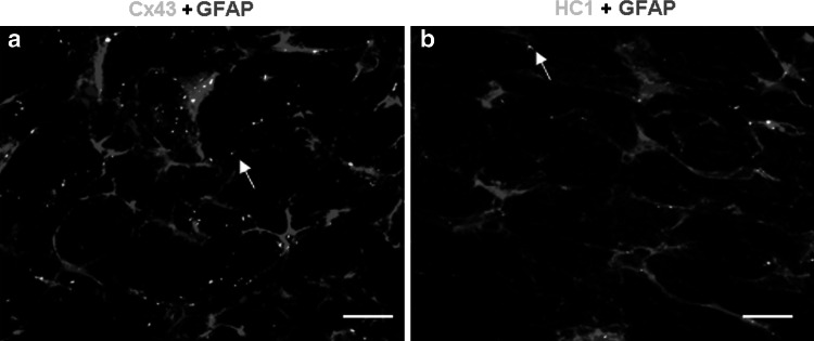
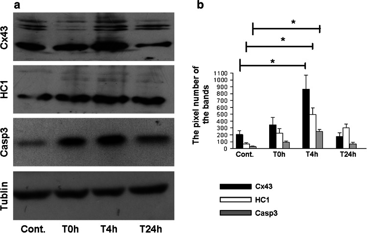
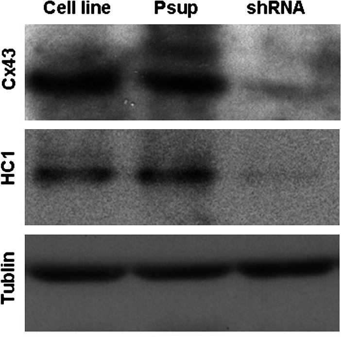
The aim of this study was to investigate the role of connexin 43 (Cx43) and its hemichannel (HC1) in the death of astrocytes following ischemia/reperfusion (IR) or oxygen-glucose deprivation/reoxygenation (OGDR) insult. Wistar rats had their bilateral common carotid artery clamped for 1.5 h followed by 0, 4, and 24 h of reperfusion (n = 8 for each time point), respectively. All rats were sacrificed and Cx43, HC1, and caspase 3 (Casp3) in cerebral ischemic tissues were examined by immunohistochemistry and western blotting. Astrocytes cell line, astrocytes transduced with a retroviral empty vector (Psup astrocyte), or a Cx43-specific shRNA construct (shRNA astrocytes) were treated with OGDR insult for various periods. The viability of astrocytes was assessed by MTT assay. The expression of Cx43, HC1, and Casp3 was detected with western blotting. The results showed that the expression of Cx43, HC1, and Casp3 in rats' brain, astrocytes, and Psup astrocytes was significantly increased after 4 h of IR/OGDR and recovered on 24 h of the insult. Cell viability decreased after 4 h of the insult whereas the cell viability increased on 24 h after the insult. In contrast, the expression of Cx43, HC1, Casp3, and cell viability had no statistical differences in the null Cx43 gene-shRNA transfected astrocytes after the treatment of OGDR. The results suggest that Cx43 and HC1 are likely to play the pivotal roles in the mediation of the astrocytic death.


Figures

Fig. 1
Fig. 1
Double immunostaining for the expression of Cx43 + GFAP, HC1 + GFAP in the astrocytes from rat’s brain after IR insult. Cx43 and HC1 were stained green, glial fibrillary acidic protein (GFAP) was stained red, and nuclei were stained blue. The arrows show the positive staining of Cx43/HC1. Bar 50 μm (Color figure online)
Fig. 2
Fig. 2
Immunostaining for the expression of Cx43, HC1, and Casp3 in rat’s brain after IR insult. Cx43, HC1, and Casp3 were stained green and nuclei were stained blue. SVZ subventricular zone, LV lateral ventricle. Bar 25 μm. a1a3 The expression of Cx43, HC1, and Casp3 in SVZ from rat without the insult of ischemia and reperfusion (Cont.). b1b3 The expression of Cx43, HC1, and Casp3 in SVZ from rat after 0 h of reperfusion (T0 h). c1c3 The expression of Cx43, HC1, and Casp3 in SVZ from rat after 4 h of reperfusion (T4 h). d1d3 The expression of Cx43, HC1, and Casp3 in SVZ from rat after 24 h of reperfusion (T24 h). e1 The plaque area (pixels) of Cx43 and HC1 staining per cell with the time points. e2 The percentage of Casp3 positive cells (%) with the time points. *P < 0.05 (Color figure online)
Fig. 3
Fig. 3
Western blot analysis of Cx43, HC1, and Casp3 expression in subventricular zone of rat’s brain after IR insult. a Protein expression of Cx43, HC1, Casp3, and tublin were identified from the tissue extracts in subventricular zone of rat’s brain. b Bar histograms show the mean ± SD corresponding to data displayed in part a. *P < 0.05
Fig. 4
Fig. 4
Western blot to confirm the effect of transfection with null Cx43 gene shRNA in astrocytes. After 24 h of the gene transfection, the lysate from astrocytes was resolved by SDS-PAGE, and immuno-positive bands were semi-quantified by scanning laser densitometry. Protein expression of Cx43, HC1, and tublin was identified, respectively
Fig. 5
Fig. 5
Viability of astrocytes after OGDR treatment. Astrocytes were cultured in 96-well plates at a density of 5,000 cells/well and treated with OGDR. Cells without the treatments of OGDR acted as a control. Cell viability was assessed by the MTT assay (MTT measured in units of optical density at 570 nm). a Cell line astrocytes, b Psup astrocytes, c shRNA astrocytes. *P < 0.05
Fig. 6
Fig. 6
Western blot analysis of Cx43, HC1, and Casp3 expression from astrocytes following OGDR treatment. Astrocytes were treated with OGDR techniques. The lysates were resolved by SDS-PAGE, and immuno-positive bands were semi-quantified by scanning laser densitometry. Data are expressed as mean ± SD corresponding to data displayed, *P < 0.05. a1a2 Astrocytes cell line. b1b2 Psup astrocytes. c1c2 shRNA astrocytes. d1 Tublin was detected as a control

References

    1. Benjelloun N, Joly LM, Palmier B, Plotkine M, Charriaut-Marlangue C (2003) Apoptotic mitochondrial pathway in neurones and astrocytes after neonatal hypoxia–ischaemia in the rat brain. Neuropathol Appl Neurobiol 29:350–360 - PubMed
    1. Bennett MV, Contreras JE, Bukauskas FF, Sáez JC (2003) New roles for astrocytes: gap junction hemichannels have something to communicate. Trends Neurosci 26:610–617 - PMC - PubMed
    1. Carr AJ, Whitmore D (2005) Imaging of single light-responsive clock cells reveals fluctuating free-running periods. Nat Cell Biol 7:319–321 - PubMed
    1. Cooper CD, Solan JL, Dolejsi MK, Lampe PD (2000) Analysis of connexin phosphorylation sites. Methods 20:196–204 - PubMed
    1. Cusato K, Bosco A, Rozental R, Guimarães CA, Reese BE, Linden R, Spray DC (2003) Gap junctions mediate bystander cell death in developing retina. J Neurosci 23:6413–6422 - PMC - PubMed

Publication types

MeSH terms

LinkOut - more resources