. 2013 Jan 18;339(6117):324-8.
doi: 10.1126/science.1231921.

GDE2 promotes neurogenesis by glycosylphosphatidylinositol-anchor cleavage of RECK

Affiliations

GDE2 promotes neurogenesis by glycosylphosphatidylinositol-anchor cleavage of RECK

Sungjin Park et al. Science. .

Abstract

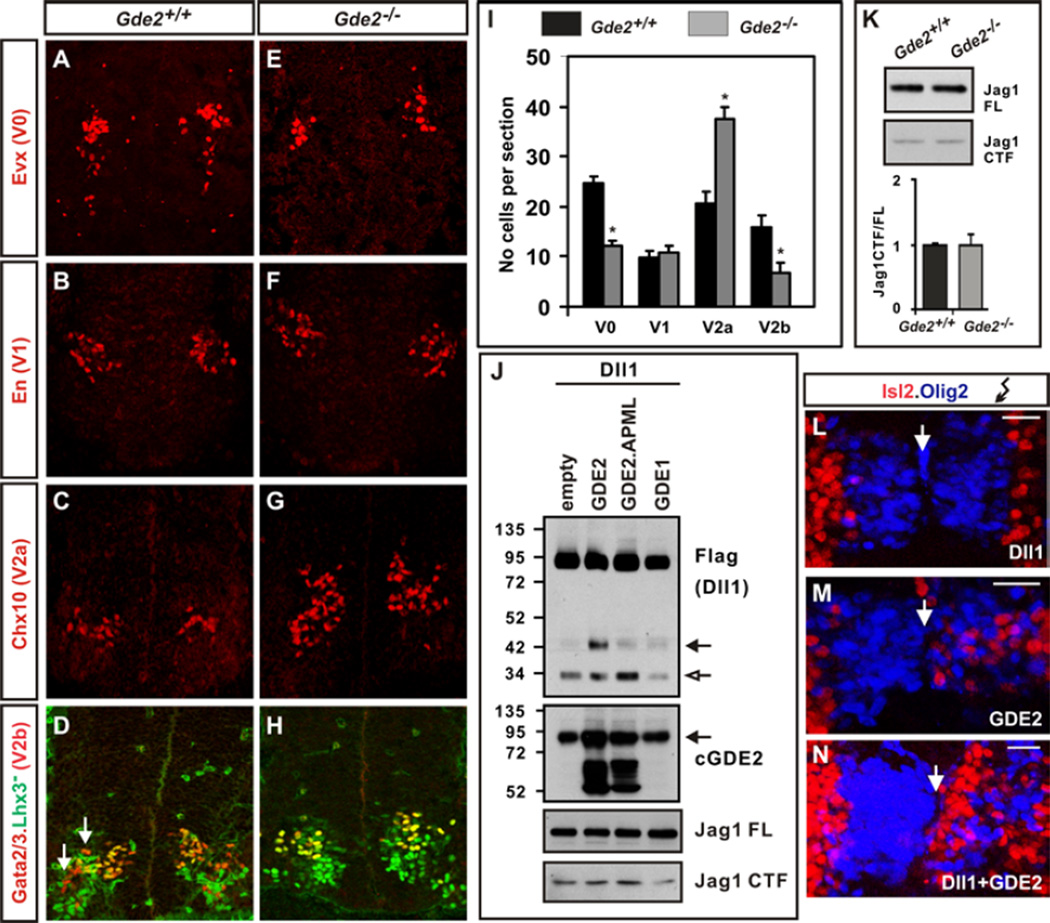
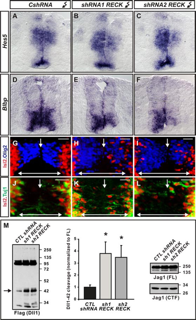
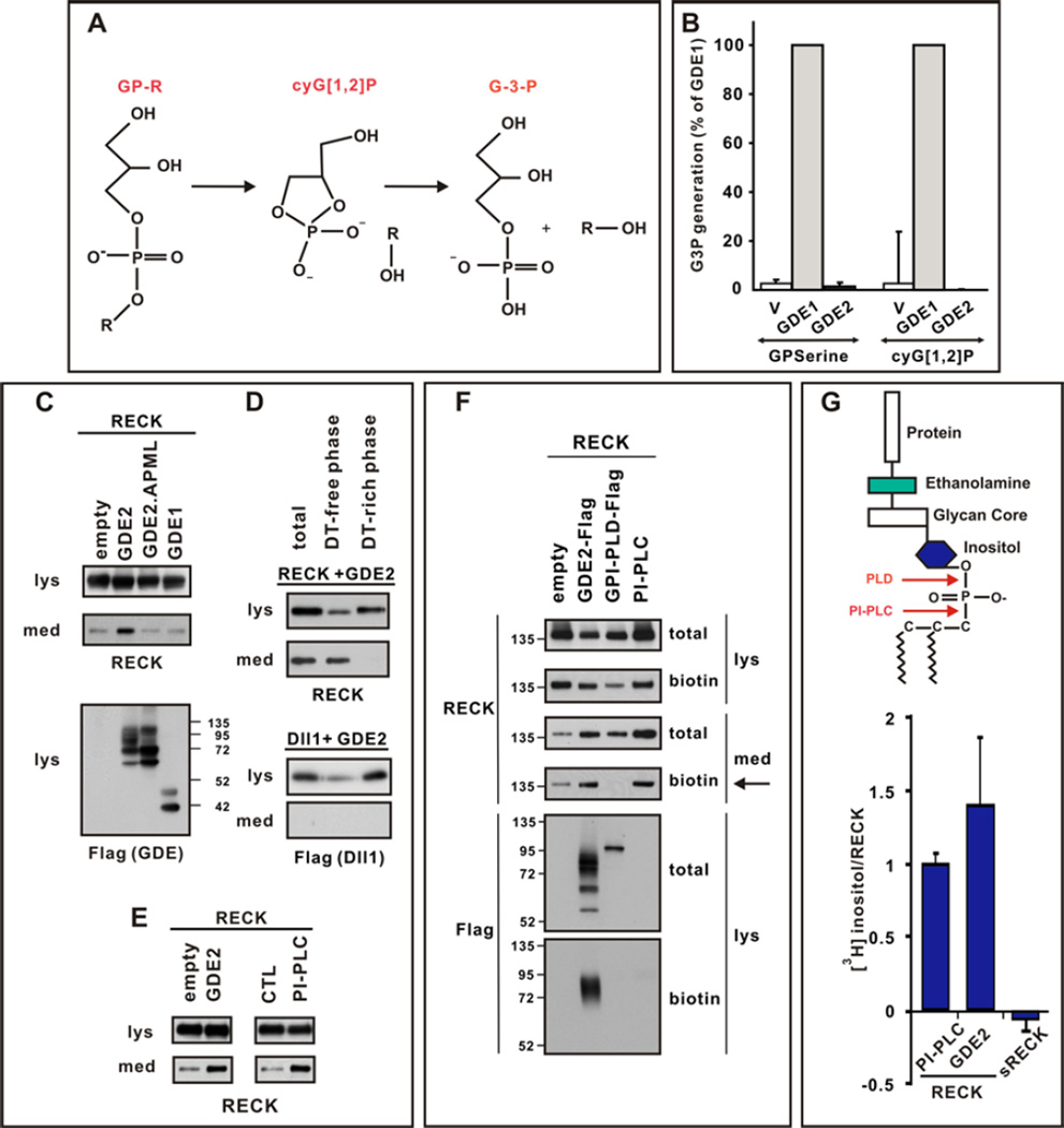
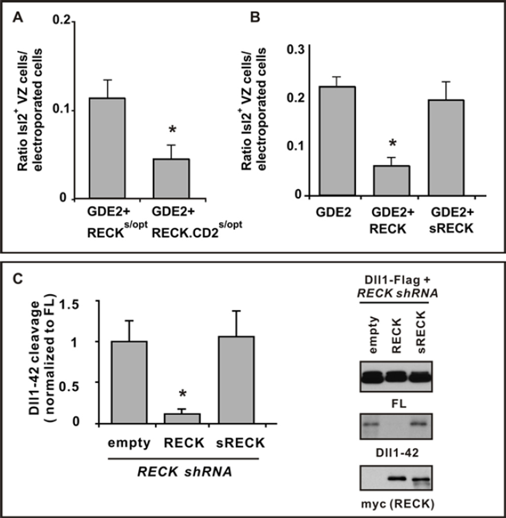
The six-transmembrane protein glycerophosphodiester phosphodiesterase 2 (GDE2) induces spinal motor neuron differentiation by inhibiting Notch signaling in adjacent motor neuron progenitors. GDE2 function requires activity of its extracellular domain that shares homology with glycerophosphodiester phosphodiesterases (GDPDs). GDPDs metabolize glycerophosphodiesters into glycerol-3-phosphate and corresponding alcohols, but whether GDE2 inhibits Notch signaling by this mechanism is unclear. Here, we show that GDE2, unlike classical GDPDs, cleaves glycosylphosphatidylinositol (GPI) anchors. GDE2 GDPD activity inactivates the Notch activator RECK (reversion-inducing cysteine-rich protein with kazal motifs) by releasing it from the membrane through GPI-anchor cleavage. RECK release disinhibits ADAM (a disintegrin and metalloproteinase) protease-dependent shedding of the Notch ligand Delta-like 1 (Dll1), leading to Notch inactivation. This study identifies a previously unrecognized mechanism to initiate neurogenesis that involves GDE2-mediated surface cleavage of GPI-anchored targets to inhibit Dll1-Notch signaling.


Figures

Figure 1
Figure 1. Stimulation of Dll1 shedding by GDE2
(A–H) Coronal sections of E13.5 mouse spinal cords. Arrows mark V2b interneurons (red). (I) Graph quantifying interneuron numbers in WT and Gde2−/− mutants; mean ± s.e.m, n= 4, two-tailed t-test: V0 *p= 0.0016; V1 p= 0.4778; V2a *p= 0.0028, V2b *p= 0.0088 (J) Western blots of extracts of chick spinal cords electroporated with Dll1-Flag plasmid; open arrow = 30kD Dll1 C-terminal fragment, black arrow = C-terminal 42kD Dll1 product (Dll1-42). Arrow (GDE2) = endogenous glycosylated GDE2, lower bands are hypoglycosylated GDE2. (K) Western blot of Jag1 processing (FL= full length; CTF= C-terminal fragment) and quantification of Jag1 CTF/FL ratios from E12.5 embryonic spinal cord extracts. (L–N) Close up of electroporated chick spinal cords (right) shows increased Isl2+ MNs (red) when Dll1 is coelecroporated with GDE2. Arrow = midline. Scale bar = 20µm.
Figure 2
Figure 2. Effects of RECK ablation
(A–F) Notch target gene mRNAs are reduced in HH st19/20 chick spinal cords electroporated with RECK shRNAs but not control CshRNAs. (G–L) Olig2 expression (blue) demarcates VZ of chick spinal cords electroporated (right) with control and RECK shRNAs, showing Isl2+ MNs (red) that express neuronal Tuj1 (green) when RECK is knocked-down. Arrow = midline; doubleheaded arrow = VZ. Scale bar = 20µm. (M) Western blots of chick spinal cords electroporated with Dll1-Flag plasmid and RECK shRNAs show RECK knockdown stimulates Dll1-42 production (arrow) but Jag1 expression and processing is unchanged. Graph quantifying Dll1-42 cleavage from Westerns; mean ± s.e.m. Two-tailed t-test, n=4, sh1RECK *p= 0.0066 sh2RECK *p=0.0175.
Figure 3
Figure 3. GDE2 cleaves RECK within the GPI-anchor
(A) Schematic of 2-step GDPD catalysis. (B) Graph quantifying in vitro GDPD assay in transfected HEK293T cells using glycerophosphoserine (GPserine) and synthetic cyclic glycerol[1,2] phosphate intermediate. (C–E) Western blots of transfected HEK293T cell lysates (lys) and medium (med). (C) RECK is detected in the medium when catalytically active GDE2 is present. (D) After sequential Triton X-114 extraction cleaved RECK is observed in the Detergent (DT)-free hydrophilic phase, while Dll1, which is not cleaved by GDE2, is retained in DT-rich hydrophobic phase of the lysate. (E) RECK ECD is generated by GDE2 or PI-PLC activities. (F) Western blot of lysates (lys) and medium (med) of HEK293T cells transfected with RECK and C-terminal Flag tagged GDE2 or GPI-PLD. Surface RECK is labeled by biotin. GDE2 but not GPI-PLD releases surface biotinylated RECK into the medium (arrow). Both GDE2 and GPI-PLD are visualized by Flag antibodies but only GDE2 is labeled by biotin indicating GDE2 is localized to the cell surface. PI-PLC was added to intact cells and serves as a positive control. (G) Schematic of GPI-anchor. Graph quantifying amount of radiolabel incorporated into RECK or secreted (s) RECK when GDE2 or PI-PLC is present. Mean ± s.e.m. n=4–12.
Figure 4
Figure 4. GDE2 inactivates RECK by GPI-anchor cleavage
(A, B) Graphs quantifying ratio of ectopic Isl2+ VZ MNs normalized to the number of transfected GDE2 cells. Mean ± s.e.m., two-tailed t-test, (A) Suboptimal levels of plasmids expressing RECKs/opt or RECK-CD2 s/opt were coelectroporated with GDE2. RECK-CD2 was more effective than RECK in suppressing GDE2 dependent MN generation, *p= 0.0306 (n=5); (B) Plasmids expressing RECK or sRECK were coelectroporated with GDE2; RECK effectively suppressed GDE2 function but sRECK did not, *p= 5.59×10⁻⁵ (n=8–10) compared with GDE2. (C) Western blot of extracts of chick spinal cords electroporated with Dll1-Flag and RECK shRNA targeting 3’ UTR to detect full-length (FL) and processed Dll1-42. The phenotype is rescued by exogenous plasmids expressing WT RECK ORF but not sRECK. Densitometric quantification of Dll1-42, mean ± s.e.m., n=4. Two-tailed t-test, *p=0.013 compared with empty.

References

    1. Rao M, Sockanathan S. Transmembrane protein GDE2 induces MN differentiation in vivo. Science. 2005;309:2212–2215. - PubMed
    1. Yan Y, Sabharwal P, Rao M, Sockanathan S. The antioxidant Prdx1 controls MN differentiation by thiol-redox dependent activation of GDE2. Cell. 2009;138:1209–1221. - PMC - PubMed
    1. Sabharwal P, Lee C, Park S, Rao M, Sockanathan S. GDE2 regulates subtype specific MN generation through inhibition of Notch signaling. Neuron. 2011;71:1058–1070. - PMC - PubMed
    1. Yanaka N. Mammalian glycerophosphodiester phosphodiesterases. Biosci. Biotechnol. Biochem. 2007;71:1811–1818. - PubMed
    1. D'Souza B, Meloty-Kapella L, Weinmaster G. Canonical and non-canonical Notch ligands. Curr. Top. Dev. Biol. 2010;92:73–129. - PMC - PubMed

Publication types

MeSH terms

Substances