

Adolescent stress-induced epigenetic control of dopaminergic neurons via glucocorticoids
- PMID: 23329051
- PMCID: PMC3617477
- DOI: 10.1126/science.1226931
Adolescent stress-induced epigenetic control of dopaminergic neurons via glucocorticoids
Abstract
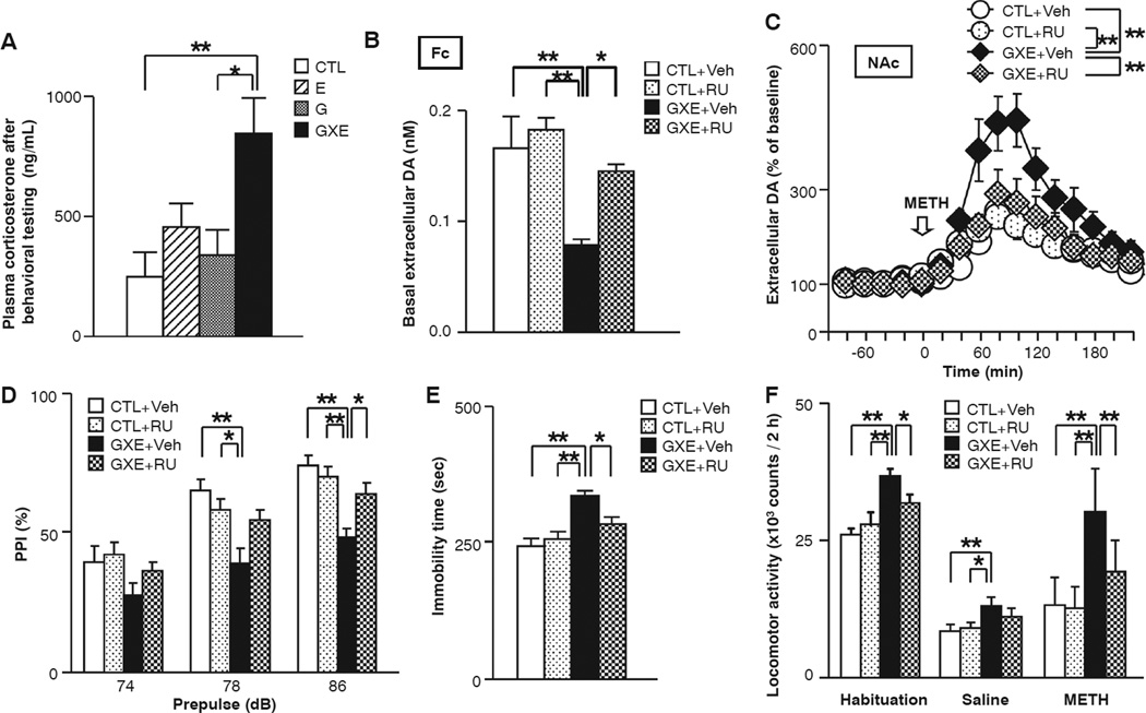
Environmental stressors during childhood and adolescence influence postnatal brain maturation and human behavioral patterns in adulthood. Accordingly, excess stressors result in adult-onset neuropsychiatric disorders. We describe an underlying mechanism in which glucocorticoids link adolescent stressors to epigenetic controls in neurons. In a mouse model of this phenomenon, a mild isolation stress affects the mesocortical projection of dopaminergic neurons in which DNA hypermethylation of the tyrosine hydroxylase gene is elicited, but only when combined with a relevant genetic risk for neuropsychiatric disorders. These molecular changes are associated with several neurochemical and behavioral deficits that occur in this mouse model, all of which are blocked by a glucocorticoid receptor antagonist. The biology and phenotypes of the mouse models resemble those of psychotic depression, a common and debilitating psychiatric disease.
Figures


Comment in
-
Neuroscience. Hormones and the social brain.Science. 2013 Jan 18;339(6117):279-80. doi: 10.1126/science.1233713. Science. 2013. PMID: 23329037 No abstract available.
-
Basic research: From stress to social behaviour-glucocorticoids and dopaminergic circuits pave the way.Nat Rev Endocrinol. 2013 Mar;9(3):125. doi: 10.1038/nrendo.2013.21. Epub 2013 Feb 5. Nat Rev Endocrinol. 2013. PMID: 23381034 No abstract available.
References
-
- Blakemore SJ. Nat. Rev. Neurosci. 2008;9:267. - PubMed
-
- Caspi A, Roberts BW, Shiner RL. Annu. Rev. Psychol. 2005;56:453. - PubMed
-
- Gunnar M, Quevedo K. Annu. Rev. Psychol. 2007;58:145. - PubMed
-
- Meaney MJ. Child Dev. 2010;81:41. - PubMed
-
- Moffitt TE, Caspi A, Rutter M. Arch. Gen. Psychiatry. 2005;62:473. - PubMed
Publication types
MeSH terms
Substances
Grants and funding
- P50 MH094268/MH/NIMH NIH HHS/United States
- MH-069853/MH/NIMH NIH HHS/United States
- MH-092443/MH/NIMH NIH HHS/United States
- R21 MH085226/MH/NIMH NIH HHS/United States
- MH-084018/MH/NIMH NIH HHS/United States
- MH-085226/MH/NIMH NIH HHS/United States
- MH-094268/MH/NIMH NIH HHS/United States
- RC1 MH088753/MH/NIMH NIH HHS/United States
- R01 MH092443/MH/NIMH NIH HHS/United States
- K99MH-094408/MH/NIMH NIH HHS/United States
- P20 MH084018/MH/NIMH NIH HHS/United States
- MH-088753/MH/NIMH NIH HHS/United States
- K99 MH094408/MH/NIMH NIH HHS/United States
- R01 MH069853/MH/NIMH NIH HHS/United States
LinkOut - more resources
Full Text Sources
Other Literature Sources
Medical
Molecular Biology Databases
