Review
. 2013 Feb;14(1):27-47.
doi: 10.1007/s40257-012-0006-4.

Diagnosis and treatment of lichen sclerosus: an update

Affiliations
Review

Diagnosis and treatment of lichen sclerosus: an update

Susanna K Fistarol et al. Am J Clin Dermatol. 2013 Feb.

Abstract

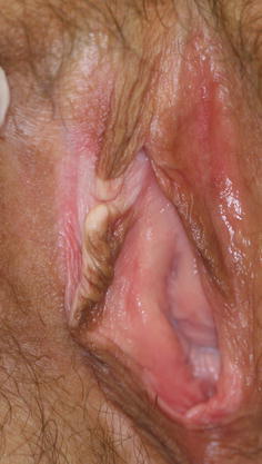
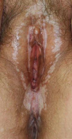
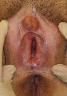
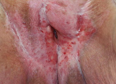
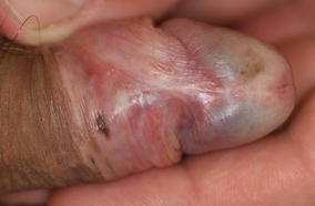
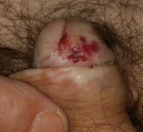
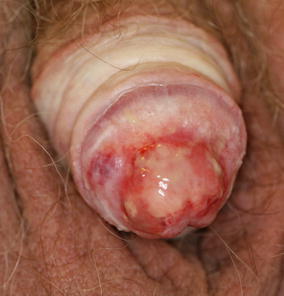
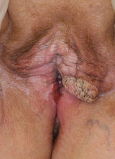
Lichen sclerosus (LS) is a chronic, inflammatory, mucocutaneous disorder of genital and extragenital skin. LS is a debilitating disease, causing itch, pain, dysuria and restriction of micturition, dyspareunia, and significant sexual dysfunction in women and men. Many findings obtained in recent years point more and more towards an autoimmune-induced disease in genetically predisposed patients and further away from an important impact of hormonal factors. Preceding infections may play a provocative part. The role for Borrelia is still controversial. Trauma and an occlusive moist environment may act as precipitating factors. Potent and ultrapotent topical corticosteroids still head the therapeutic armamentarium. Topical calcineurin inhibitors are discussed as alternatives in the treatment of LS in patients who have failed therapy with ultrapotent corticosteroids, or who have a contraindication for the use of corticosteroids. Topical and systemic retinoids may be useful in selected cases. Phototherapy for extragenital LS and photodynamic therapy for genital LS may be therapeutic options in rare cases refractory to the already mentioned treatment. Surgery is restricted to scarring processes leading to functional impairment. In men, circumcision is effective in the majority of cases, but recurrences are well described. Anogenital LS is associated with an increased risk for squamous cell carcinoma of the vulva or penis. This review updates the epidemiology, clinical presentation, histopathology, pathogenesis, and management of LS of the female and male genitals and extragenital LS in adults and children.


Figures

Fig. 1
Fig. 1
A 41-year-old woman with asymmetric vulvar lichen sclerosus limited to the upper part of the right labium minus and the interlabial sulcus: depigmentation, hyperpigmentation, and sclerosis leading to circumscribed retraction of the right labium minus
Fig. 2
Fig. 2
An 80-year-old woman with extensive, long-standing, anogenital lichen sclerosus affecting the entire vulva, perineum, perianal region, genitocrural folds, inner thighs, and buttocks: depigmentation, hyperpigmentation, erythema, sclerosis, erosions, telangiectasias, and complete loss of labia minora
Fig. 3
Fig. 3
A 65-year-old woman with lichen sclerosus showing the characteristic ‘figure-of-eight’ distribution: typical primary lesions are the ivory-white, flat, polygonal papules, giving lichen sclerosus the name ‘white-spot-disease’ in the past. Note the involvement of the interlabial sulci, clitoris, and clitorial hood
Fig. 4
Fig. 4
A 17-year-old woman with lichen sclerosus affecting primarily the periclitorial region: the periclitorial hood is sealed and the clitoris buried. Fissure in the upper part of the periclitorial hood and diffuse erythema of the vestibule
Fig. 5
Fig. 5
A 27-year-old woman with lichen sclerosus affecting the vulva and perineum: perineal pallor, sclerosis, and sagittal fissure
Fig. 6
Fig. 6
A 47-year-old woman with vulvar lichen sclerosus: diffuse pallor, erythema, sclerosis, and telangiectasias, almost complete loss of labia minora and smegmatic pseudocyst resulting from adhesions of the clitorial hood
Fig. 7
Fig. 7
A 75-year-old woman with lichen sclerosus/lichen planus overlap syndrome: diffuse pallor, erythema, sclerosis, and telangiectasias. Damage of the normal architecture with partial loss of labia minora. Edema of the clitoris and the residual labia minora. Introital erosions and narrowing of the introitus. Sagittal fissuring of the perineum. Coincidental finding is an urethral caruncle
Fig. 8
Fig. 8
An 81-year-old woman with lichen sclerosus with superimposed lichen simplex: marked lichenification and excoriations, ecchymoses, telangiectasias, erosions, and ulceration on a hypopigmented and erythematous background. Fissuring of the right genitocrural fold. Complete loss of normal vulvar architecture
Fig. 9
Fig. 9
A 6-year-old girl with anogenital lichen sclerosus: slight sclerosis of the clitorial hood and the inner aspect of labia majora. Prominent contusiform hemorrhage
Fig. 10
Fig. 10
A 37-year-old man with genital lichen sclerosus: grayish-bluish, white discoloration of the glans and inner surface of the prepuce, sclerosis of the frenulum, and freckled melanosis
Fig. 11
Fig. 11
A 65-year-old man with genital lichen sclerosus: depigmentation, macular erythema, and severe sclerosis leading to phimosis
Fig. 12
Fig. 12
A 57-year-old man with genital lichen sclerosus: depigmentation, atrophy of the glans with crinkling appearance, ecchymoses, and macular melanosis
Fig. 13
Fig. 13
A 73-year-old man with genital lichen sclerosis: severe sclerosis, scarring of the coronal sulcus, and large ulceration of the glans
Fig. 14
Fig. 14
A 65-year-old woman (same patient as in Fig. 3) with genital and extragenital lichen sclerosus on the volar aspects of the wrists: characteristic bluish-white, polygonal, slightly elevated papules coalescing into plaques
Fig. 15
Fig. 15
An 81-year-old woman with genital and extragenital lichen sclerosus on the upper thighs: papular, partially atrophic lesion with wrinkled surface, telangiectases, and hemorrhage
Fig. 16
Fig. 16
A 65-year-old woman with extragenital LS on the volar aspect of the wrist (same patient as in Fig. 3 and Fig. 14) with prominent follicular plugging
Fig. 17
Fig. 17
An 89-year-old woman with long-standing anogenital lichen sclerosus. She developed a nodular lesion firm to palpation on the left side of the vulva. Biopsy confirmed an invasive carcinoma

Similar articles

Cited by

References

    1. Wallace HJ. Lichen sclerosus et atrophicus. Trans St Johns Hosp Dermatol Soc. 1971;57:9–30. - PubMed
    1. Brown AR, Dunlap CL, Bussard DA, et al. Lichen sclerosus et atrophicus of the oral cavity: report of two cases. Oral Surg Oral Med Oral Pathol Oral Radiol Endod. 1997;84:165–170. - PubMed
    1. Jensen T, Worsaae N, Melgaard B. Oral lichen sclerosus et atrophicus: a case report. Oral Surg Oral Med Oral Pathol Oral Radiol Endod. 2002;94:702–706. - PubMed
    1. Azevedo RS, Romañach MJ, de Almeida OP, et al. Lichen sclerosus of the oral mucosa: clinicopathological features of six cases. Int J Oral Maxillofac Surg. 2009;38:855–860. - PubMed
    1. Neill SM, Lewis FM, Tatnall FM, et al. British Association of Dermatologists’ guidelines for the management of lichen sclerosus 2010. Br J Dermatol. 2010;163:672–682. - PubMed

MeSH terms