

Fabrication of two-layer poly(dimethyl siloxane) devices for hydrodynamic cell trapping and exocytosis measurement with integrated indium tin oxide microelectrodes arrays
- PMID: 23329291
- PMCID: PMC5002351
- DOI: 10.1007/s10544-013-9744-1
Fabrication of two-layer poly(dimethyl siloxane) devices for hydrodynamic cell trapping and exocytosis measurement with integrated indium tin oxide microelectrodes arrays
Abstract
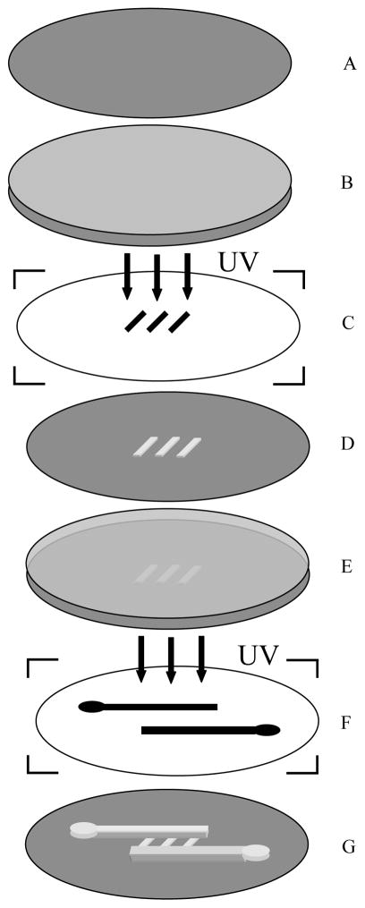
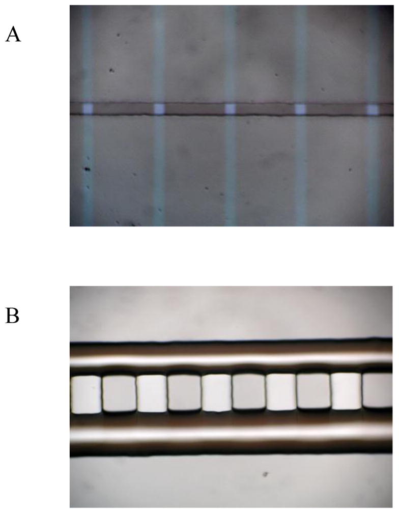
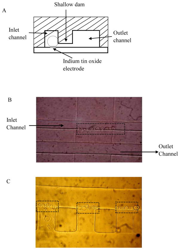
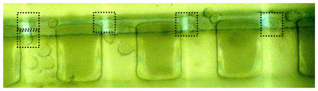
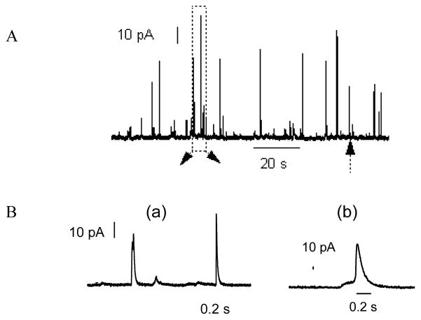
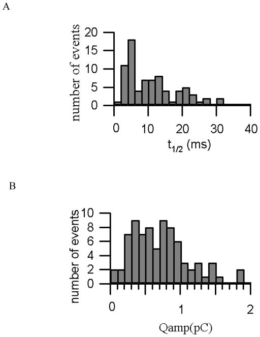
The design, fabrication and test of a microfluidic cell trapping device to measure single cell exocytosis were reported. Procedures on the patterning of double layer template based on repetitive standard photolithography of AZ photoresist were investigated. The replicated poly(dimethyl siloxane) devices with 2.5 μm deep channels were proved to be efficient for stopping cells. Quantal exocytosis measurement can be achieved by targeting single or small clumps of chromaffin cells on top of the 10 μm × 10 μm indium tin oxide microelectrodes arrays with the developed microdevice. And about 72 % of the trapping sites can be occupied by cells with hydrodynamic trapping method and the recorded amperometric signals are comparable to the results with traditional carbon fiber microelectrodes. The method of manufacturing the microdevices is simple, low-cost and easy to perform. The manufactured device offers a platform for the high throughput detection of quantal catecholamine exocytosis from chromaffin cells with sufficient sensitivity and broad application.
Figures
Similar articles
-
On-chip amperometric measurement of quantal catecholamine release using transparent indium tin oxide electrodes.Anal Chem. 2006 Apr 15;78(8):2521-5. doi: 10.1021/ac052037d. Anal Chem. 2006. PMID: 16615759
-
Controlled on-chip stimulation of quantal catecholamine release from chromaffin cells using photolysis of caged Ca2+ on transparent indium-tin-oxide microchip electrodes.Lab Chip. 2008 Jan;8(1):161-9. doi: 10.1039/b715308m. Epub 2007 Oct 26. Lab Chip. 2008. PMID: 18094774 Free PMC article.
-
Magnetron sputtered diamond-like carbon microelectrodes for on-chip measurement of quantal catecholamine release from cells.Biomed Microdevices. 2008 Oct;10(5):623-9. doi: 10.1007/s10544-008-9173-8. Biomed Microdevices. 2008. PMID: 18493856 Free PMC article.
-
Electrochemical measurement of quantal exocytosis using microchips.Pflugers Arch. 2018 Jan;470(1):97-112. doi: 10.1007/s00424-017-2063-2. Epub 2017 Sep 2. Pflugers Arch. 2018. PMID: 28866728 Free PMC article. Review.
-
Tunable Microfluidic Devices for Hydrodynamic Fractionation of Cells and Beads: A Review.Sensors (Basel). 2015 Nov 24;15(11):29685-701. doi: 10.3390/s151129685. Sensors (Basel). 2015. PMID: 26610519 Free PMC article. Review.
Cited by
-
On-Chip Cyclic Voltammetry Measurements Using a Compact 1024-Electrode CMOS IC.Anal Chem. 2021 Jun 8;93(22):8027-8034. doi: 10.1021/acs.analchem.1c01132. Epub 2021 May 26. Anal Chem. 2021. PMID: 34038637 Free PMC article.
-
Instrumented Microphysiological Systems for Real-Time Measurement and Manipulation of Cellular Electrochemical Processes.iScience. 2019 Nov 22;21:521-548. doi: 10.1016/j.isci.2019.10.052. Epub 2019 Oct 28. iScience. 2019. PMID: 31715497 Free PMC article. Review.
-
Surface-modified CMOS IC electrochemical sensor array targeting single chromaffin cells for highly parallel amperometry measurements.Pflugers Arch. 2018 Jan;470(1):113-123. doi: 10.1007/s00424-017-2067-y. Epub 2017 Sep 9. Pflugers Arch. 2018. PMID: 28889250 Free PMC article. Review.
-
Large-Area and High-Throughput PDMS Microfluidic Chip Fabrication Assisted by Vacuum Airbag Laminator.Micromachines (Basel). 2017 Jul 12;8(7):218. doi: 10.3390/mi8070218. Micromachines (Basel). 2017. PMID: 30400409 Free PMC article.
-
A microfluidic platform for chemical stimulation and real time analysis of catecholamine secretion from neuroendocrine cells.Lab Chip. 2013 Dec 7;13(23):4663-73. doi: 10.1039/c3lc50779c. Lab Chip. 2013. PMID: 24126415 Free PMC article.
References
Publication types
MeSH terms
Substances
Grants and funding
LinkOut - more resources
Full Text Sources
Other Literature Sources
