. 2013 Feb;35(2):151-7.
doi: 10.1007/s10059-013-2298-5. Epub 2013 Jan 16.

Berberine protects 6-hydroxydopamine-induced human dopaminergic neuronal cell death through the induction of heme oxygenase-1

Affiliations

Berberine protects 6-hydroxydopamine-induced human dopaminergic neuronal cell death through the induction of heme oxygenase-1

Jinbum Bae et al. Mol Cells. 2013 Feb.

Abstract

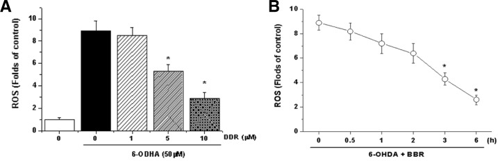
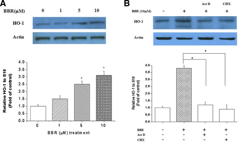
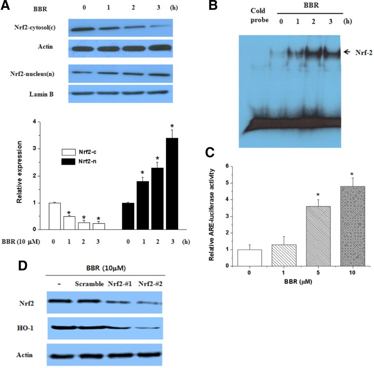
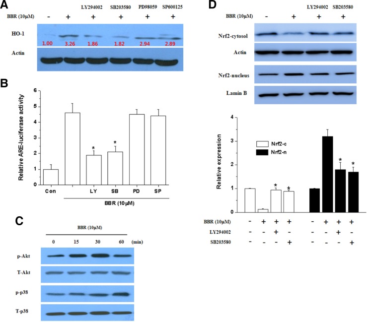
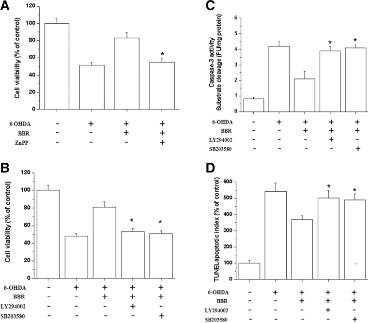
Berberine (BBR) is one of the major alkaloids and has been reported to have a variety of pharmacologic effects, including inhibition of cell cycle progression. Here, we investigated the mechanisms of BBR protection of neuronal cells from cell death induced by the Parkinson's disease-related neurotoxin 6-hydroxydopamine (6-OHDA). Pretreatment of SH-SY5Y cells with BBR significantly reduced 6-OHDAinduced generation of reactive oxygen species (ROS), caspase-3 activation, and subsequent cell death. BBR also upregulated heme oxygenase-1 (HO-1) expression, which conferred protection against 6-OHDA-induced dopaminergic neuron injury and besides, effect of BBR on HO-1 was reversed by siRNA-Nrf2. Furthermore, BBR induced PI3K/Akt and p38 activation, which are involved in the induction of Nrf2 expression and neuroprotection. These results suggest that BBR may be useful as a therapeutic agent for the treatment of dopaminergic neuronal diseases.


Figures

Fig. 1.
Fig. 1.
BBR prevents 6-OHDA-induced cell death. SH-SY5Y cells were treated with BBR (1, 5, and 10 μM) for 12 h then incubated with 6-OHDA (50 or 100 μM) for an additional 24 h. (A) Cell viability was measured by MTT assays. (B) TUNEL apoptotic index were performed according to the manufacturer’s instruction. (C) The catalytic activities of caspase-3 in cell lysates were assayed using the specific substrates DEVD-pNA. *Significantly different from BBR plus 6-OHDA-treated cells.
Fig. 2.
Fig. 2.
Effects of BBR on 6-OHDA-induced cellular ROS production. (A) Cells were pretreated with BBR (1–10 μM) or vehicle for 12 h. After the medium was removed, SH-SY5Y cells were exposed to 6-OHDA (50 μM) for 6 h and then fluorescence was measured as described in the “Materials and Methods”. (B) Prevention of 6-OHDA-induced ROS by BBR depends on the duration of BBR pretreatment. *Significantly different from BBR plus 6-OHDA-treated cells.
Fig. 3.
Fig. 3.
Effect of BBR on HO-1 mRNA and protein expression. (A) Cells were exposed to various concentrations of BBR for 12 h and total RNA was extracted. HO-1 mRNA and protein expression were analyzed by RT-PCR and Western blotting. (B) Cells were either untreated or pretreated with 50 μM cycloheximide or 10 μg/ml actinomycin D for 2 h prior to the addition of 10 μM BBR for an additional 12 h. (C) Cells were transfected with scramble (siRNA) or siNrf2 as described in the “Materials and Methods”. After 24 h incubation with BBR (10 μM), cells were harvested and subjected to Western blot for HO-1 expression. Transfection efficiency was confirmed by checking Nrf2 expression.
Fig. 4.
Fig. 4.
Effect of BBR on Nrf2 translocation into the nucleus and its transcriptional activity. (A) Effect of BBR on translocation of Nrf2. SH-SY5Y cells were treated with BBR (10 μM) for the indicated time and then nuclear and cytosolic extracts were prepared for western blotting. The Nrf2 protein level was normalized using lamin B (nuclear) and actin (cytosol). Each blot is representative of three independent experiments. (B) Effect of BBR on ARE-binding activity of Nrf2. Nuclear extracts that were prepared from cells were treated with 10 μM of BBR for the indicated times. (C) Effect of BBR on the transcriptional activity of HO-1. Cells were transfected with the ARE reporter plasmid and treated with BBR (1–10 μM). After 18 h of treatment, luciferase activities were determined. *Significantly different from BBR plus 6-OHDA-treated cells.
Fig. 5.
Fig. 5.
Induction of HO-1 and activation of Nrf2 by BBR via the PI3K/Akt and p38 pathways. (A) Effect of BBR on the phosphorylation of Akt and p38. Parallel immunoblots were analyzed for total kinase levels with anti-Akt, ERK1/2, p38 and JNK1/2 antibodies. (B) Effect of PI3K and p38 inhibitors on BBR-induced HO-1 expression. Whole cell lysates were subjected to Western blotting analysis by using anti-HO-1 and anti-actin antibodies. (C) Effect of PI3K and p38 inhibitors on BBR-induced Nrf2 translocation. Nuclear and cytosolic extracts were subjected to Western blot analysis using anti-Nrf2, anti-actin and anti-lamin B antibodies. The Nrf2 protein level was normalized using lamin B (nuclear) and actin (cytosol). Each blot is representative of three independent experiments. (D) Effect of PI3K and p38 inhibitors on BBR-induced ARE-luciferase activity. The luciferase activity was measured by luminometer. *Significantly different from BBR plus 6-OHDA-treated cells.
Fig. 6.
Fig. 6.
PI3K/Akt and p38 inhibitors atenuate the anti-apoptotic effect of BBR. (A) The HO-1 enzyme inhibitor ZnPP reversed the protective effect of BBR against 6-OHDA-induced cell death. SH-SY5Y cells were treated with BBR (10 μM) for 12 h and then incubated with different doses of ZnPP (5 μM) for 30 min; 6-OHDA (50 μM) was then added to the cells for an additional 24 h. Cell viability was detected by the MTT assay. (B) SH-SY5Y cells were pretreated for 30 min with LY294002 and SB203580 prior to the addition of BBR. Following a 12 h incubation with BBR, cells were treated with 6-OHDA for 24 h. Cell viability was assessed by the MTT assay after 6-OHDA treatment. (C) The catalytic activity of caspase-3 in cell lysates was assayed using the specific substrate DEVD-pNA. (D) TUNEL apoptotic index was performed as described in the materials and methods section. * Significantly different from BBR plus 6-OHDA-treated cells.

References

    1. Alam J., Cook J.L. Transcriptional regulation of the heme oxygenase-1 gene via the stress response element pathway. Curr. Pharm. Des. 2003;9:2499–2511. - PubMed
    1. Alam J., Stewart D., Touchard C., Boinapally S., Choi A.M., Cook J.L. Nrf2, a Cap‘n’Collar transcription factor, regulates induction of the heme oxygenase-1 gene. J. Biol. Chem. 1999;274:26071–26078. - PubMed
    1. Balogun E., Hoque M., Gong P., Killeen E., Green C.J., Foresti R., Alam J., Motterlini R. Curcumin activates the haem oxygenase-1 gene via regulation of Nrf2 and the antioxidant-responsive element. Biochem. J. 2003;371:887–895. - PMC - PubMed
    1. Chen X.L., Kunsch C. Induction of cytoprotective genes through Nrf2/antioxidant response element pathway: a new therapeutic approach for the treatment of inflammatory diseases. Curr. Pharm. Des. 2004;10:879–891. - PubMed
    1. Chen K., Gunter K., Maines M.D. Neurons overexpressing heme oxygenase-1 resist oxidative stress-mediated cell death. J. Neurochem. 2000;75:304–313. - PubMed

Publication types

MeSH terms

LinkOut - more resources