Review
. 2013 Mar;91(3):311-21.
doi: 10.1007/s00109-013-0993-5. Epub 2013 Jan 18.

Ocular neovascularization

Affiliations
Review

Ocular neovascularization

Peter A Campochiaro. J Mol Med (Berl). 2013 Mar.

Abstract

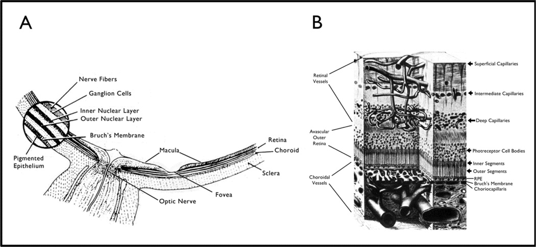
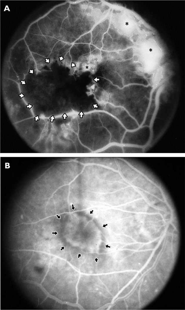
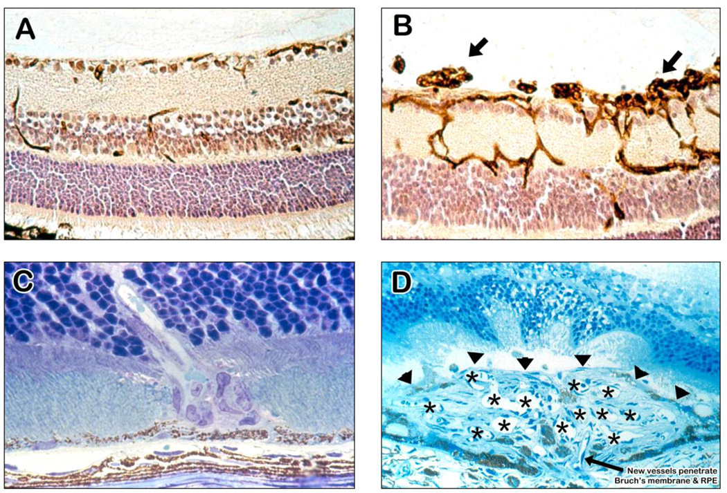
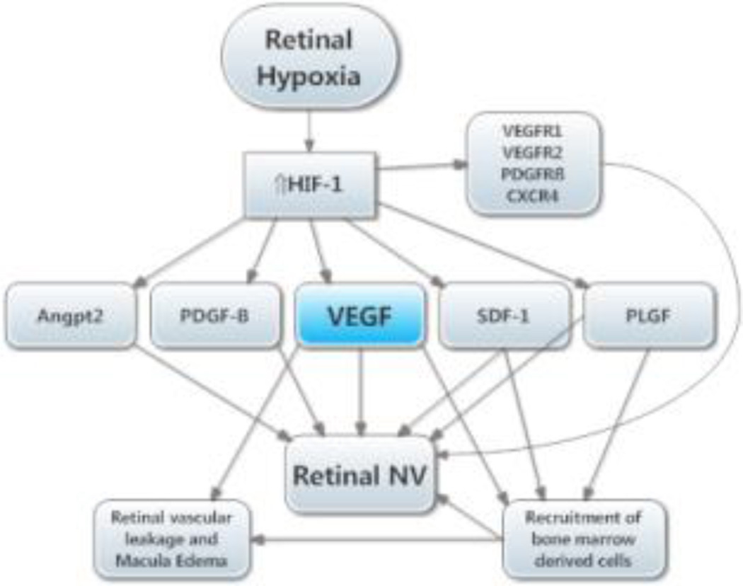
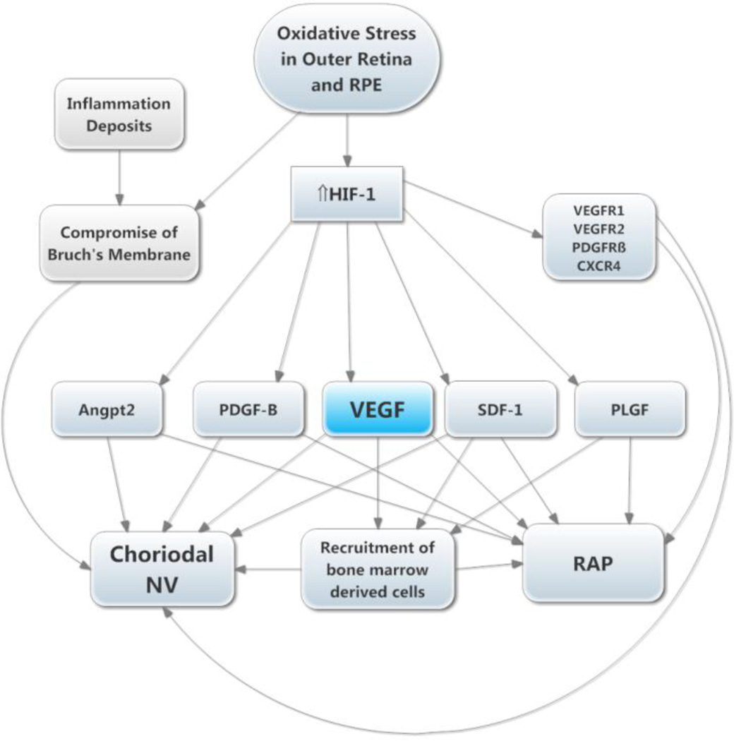
Retinal and choroidal vascular diseases constitute the most common causes of moderate and severe vision loss in developed countries. They can be divided into retinal vascular diseases, in which there is leakage and/or neovascularization (NV) from retinal vessels, and subretinal NV, in which new vessels grow into the normally avascular outer retina and subretinal space. The first category of diseases includes diabetic retinopathy, retinal vein occlusions, and retinopathy of prematurity, and the second category includes neovascular age-related macular degeneration (AMD), ocular histoplasmosis, pathologic myopia, and other related diseases. Retinal hypoxia is a key feature of the first category of diseases resulting in elevated levels of hypoxia-inducible factor-1 (HIF-1) which stimulates expression of vascular endothelial growth factor (VEGF), platelet-derived growth factor-B (PDGF-B), placental growth factor, stromal-derived growth factor-1 and their receptors, as well as other hypoxia-regulated gene products such as angiopoietin-2. Although hypoxia has not been demonstrated as part of the second category of diseases, HIF-1 is elevated and thus the same group of hypoxia-regulated gene products plays a role. Clinical trials have shown that VEGF antagonists provide major benefits for patients with subretinal NV due to AMD and even greater benefits are seen by combining antagonists of VEGF and PDGF-B. It is likely that addition of antagonists of other agents listed above will be tested in the future. Other appealing strategies are to directly target HIF-1 or to use gene transfer to express endogenous or engineered anti-angiogenic proteins. While substantial progress has been made, the future looks even brighter for patients with retinal and choroidal vascular diseases.


Figures

Figure 1
Figure 1. Location of the retina within the eye (A) and retinal blood vessels within the retina (B)
(A) Schematic cross-section of the posterior part of the eye around the optic nerve shows the macula which is located temporal to the optic nerve and is responsible for our best vision. The foveal pit or depression is located in the center of the macula and is the area needed for the very best vision. The round blow up on the left shows that the multilayered retina sits on the retinal pigmented epithelium (RPE) which sits on Bruch’s membrane. The central retinal artery enters through the optic nerve, sends numerous branches along the surface of the retina to the peripheral edges of the retina. The arterioles enter capillaries which enter venules and then veins that run along the surface of the retina and enter the central retinal vein that exits through the optic nerve. (B) A further blow up of the retina shows that the retinal arteries branch to form the superficial capillary bed near the surface of the retina and send penetrating branches to form the intermediate and deep capillaries. The outer third of the retina which consists of the photoreceptor outer and inner segments and cells bodies is avascular. It receives oxygen and nutrients from the choroidal circulation. Large choroidal vessels branch and become progressively smaller until they form the choriocapillaris which is fenestrated and allows plasma to pool along Bruch’s membrane. The RPE, which has barrier characteristics prevents fluid from entering the outer retina but allows oxygen and nutrients to enter.
Figure 2
Figure 2. Fluorescein angiograms from patients with proliferative diabetic retinopathy, an ischemic retinopathy (A), or subretinal neovascularization (B)
Sodium fluorescein was injected intravenously and then a fundus camera with filters that block extraneous wavelengths of light was used to record the image caused by emission of fluorescence from the retina. The white dye should be confined within retinal vessels, which progressively branch to form capillaries. (A) In a patient with proliferative diabetic retinopathy, the major retinal vessels branch into smaller vessels that extend into the macula. The resolution is not sufficient to see individual capillaries; they merge together and appear as gray areas between large vessels. There is a black area (surrounded by white arrows) of nonperfused retina in which all of the capillaries and larger vessels are closed due to damage from diabetes. There is retinal neovascularization (NV) on the surface of the retina adjacent to the nonperfused retina, at the optic disc, and above the optic disc (asterisks). The individual new vessels cannot be seen because they have leaked dye into the extracellular space causing confluent white patches. (B) In a patient with choroidal NV due to age-related macular degeneration, major retinal vessels are seen extending from the optic nerve (far right of image) in an arc around the macula which is in the center with many branches from the arcade vessels extending into the macula. In the center of the macula an oval hyperfluorescent area is seen (arrows); it is beneath the retina and the retinal vessels pass over it. This is an area of choroidal NV which is a convoluted network of vessels like those shown histologically in Figure 3(D) viewed through the retina. Individual vessels are not seen because the vessels leak dye into the extracellular space and therefore the network of new vessels are seen as a hazy area of hyperfluorescence with a fairly distinct border.
Figure 3
Figure 3. Ocular sections from mouse models of ocular neovascularization (NV)
(A) A normal mouse retina histochemically stained with a lectin that selectively stains vascular cells and counter-stained to show other retinal cells illustrates the 3 capillary beds of the retina with some penetrating vessels showing connections or part of connections between the beds. (B) A retina from a mouse with oxygen-induced ischemic retinopathy shows dilation of the vessels within the retina with NV on the surface of the retina (arrows). (C) A retina from a rho/VEGF transgenic mouse, a model of retinal angiomatous proliferation, shows a vessel extending from the deep capillary bed of the retina through the photoreceptors into the subretinal space. (D) A retina from a mouse with choroidal NV after laser-induced rupture of Bruch’s membrane. The arrow shows a vessel extending from the choroid through the rupture in Bruch’s membrane into the subretinal space where it connects to a large convoluted network of new vessels, many of which are cut in cross section to show their lumens (astericks). The superior margin of the choroidal NV that borders the photoreceptors is shown by the arrowheads.
Figure 4
Figure 4. Molecular pathogenesis of retinal neovascularization (NV)
This simplified version of the molecular pathogenesis of retinal NV that illustrates several important molecular signals. It highlights soluble mediators and omits cell-cell and cell-matrix signaling. Retinal NV occurs in diabetic retinopathy and other ischemic retinopathies. The underlying disease process (e.g. high glucose in diabetic retinopathy) damages retinal vessels causing vessel closure and retinal ischemia, which results in elevated HIF-1 levels. HIF-1 upregulates several vasoactive gene products including vascular endothelial growth factor (VEGF), platelet-derived growth factor (PDGF-B), placental growth factor (PLGF), stromal-derived growth factor (SDF-1), and their receptors, and angiopoietin 2 (Angpt2). VEGF causes vascular leakage and in combination with Angpt2 causes sprouting of new vessels. VEGF, SDF-1, and PLGF recruit bone marrow-derived cells which provide paracrine stimulation. PDGF-B recruits pericytes which also provide paracrine stimulation.
Figure 5
Figure 5. Molecular pathogenesis of subretinal neovascularization (NV)
This schematic highlights several important molecular signals involved in subretinal NV. Oxidative stress in the retinal pigmented epithelium (RPE) and photoreceptors causes increased levels of HIF-1, which upregulates vasoactive gene products as described above. Retinal angiomatous proliferation (RAP) occurs if VEGF levels in photoreceptors are sufficiently high to cause an adequate gradient that reaches to the deep capillary bed of the retina. Choroidal NV occurs if there is elevation of VEGF and Angpt2 combined with perturbation of Bruch’s membrane and the RPE. The other HIF-1-responsive gene products fuel the process similar to the situation in retinal NV.

References

    1. Michaelson I. The mode of development of the vascular system of the retina with some observations on its significance for certain retinal diseases. Trans Ophthalmol Soc UK. 1948;68:137–180.
    1. Baird A, Esch F, Gospodarowicz D, Guillemin R. Retina- and eye-derived endothelial cell growth factors: partial molecular chariacterization and identity with acidic and basic fibroblast growth factors. Biochemistry. 1985;24:7855–7860. - PubMed
    1. Shweiki D, Itin A, Soffer D, Keshet E. Vascular endothelial growth factor induced by hypoxia may mediate hypoxia-initiated angiogenesis. Nature. 1992;359:843–845. - PubMed
    1. Plate KH, Breier G, Welch HA, Risau W. Vascular endothelial growth factor is a potential tumor angiogenesis factor in human gliomas in vivo. Nature. 1992;359:845–848. - PubMed
    1. Aiello LP, Avery RL, Arrigg PG, Keyt BA, Jampel HD, Shah ST, Pasquale LR, Thieme H, Iwamoto MA, Park JE, et al. Vascular endothelial growth factor in ocular fluid of patients with diabetic retinopathy and other retinal disorders. N Engl J Med. 1994;331:1480–1487. - PubMed

MeSH terms