

Review
doi: 10.1074/jbc.R112.418558.
Epub 2013 Jan 17.
Mucin-type O-glycosylation during development
Affiliations
- PMID: 23329828
- PMCID: PMC3591602
- DOI: 10.1074/jbc.R112.418558
Item in Clipboard
Review
Mucin-type O-glycosylation during development
J Biol Chem.
.
Abstract
Mucin-type O-glycosylation is an evolutionarily conserved protein modification present on membrane-bound and secreted proteins. Aberrations in O-glycosylation are responsible for certain human diseases and are associated with disease risk factors. Recent studies have demonstrated essential roles for mucin-type O-glycosylation in protein secretion, stability, processing, and function. Here, we summarize our current understanding of the diverse roles of mucin-type O-glycosylation during eukaryotic development. Appreciating how this conserved modification operates in developmental processes will provide insight into its roles in human disease and disease susceptibilities.
Figures
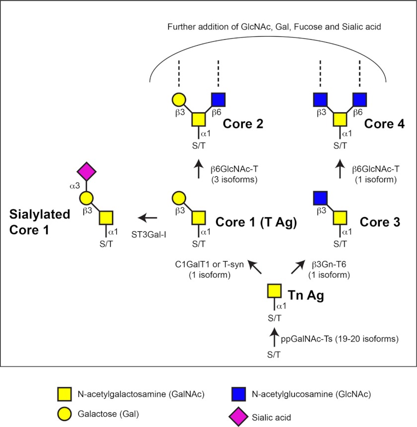
Biosynthesis of mucin-type O-glycans. The initiation of mucin-type O-glycosylation is catalyzed by the addition of GalNAc to the hydroxyl groups of serine or threonine in protein substrates destined to be membrane-bound or secreted, forming the Tn antigen (Tn Ag). The addition of other sugars results in the formation of the core structures. Enzymes responsible for the synthesis of the Tn antigen, core 1 (T antigen (T Ag)), core 2, core 3, core 4, and sialylated core 1 structures are shown. The number of isoforms present in mammals is shown in parentheses. Additional extensions of O-glycans are not shown. C1GalT1 or T-syn, core 1 β1,3-galactosyltransferase; β3Gn-T6, β1,3-N-acetylglucosaminyltransferase 6; β6GlcNAc-T, β1,6-N-acetylglucosaminyltransferase; ST3Gal-I, α2,3-sialyltransferase I.
Mucin-type O-glycan expression during development.
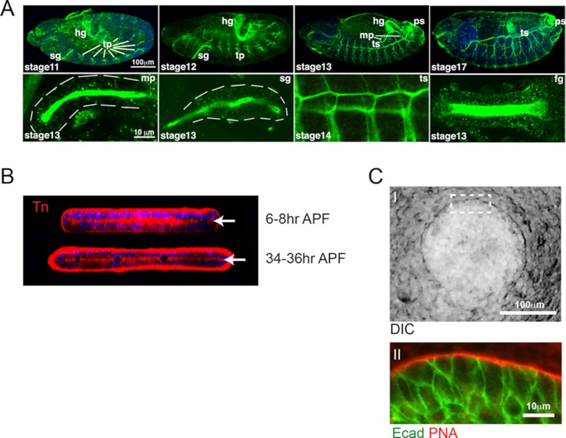
A, Tn antigen (GalNAcα1-Ser/Thr) expression during Drosophila embryonic development (as detected by the anti-Tn antibody, green). The upper panels show whole embryos at different stages of development (stages 11–13 and 17), with the salivary glands (sg), hindgut (hg), tracheal placodes (tp), tracheal system (ts), Malpighian tubules (mp), and posterior spiracles (ps) indicated. The lower panels show magnified views of the Malpighian tubules, salivary glands, tracheal placodes, and foregut (fg). The dashed white lines outline the outer edges of specific organs. This figure is adapted from Ref. . B, Tn antigen expression in Drosophila pupal wings at 6–8 and 34–36 h after puparium formation (APF). X-Z optical sections are shown and demonstrate expression of Tn antigen structures at the interface (white arrows) of the dorsal and ventral cell layers of the developing wing. This figure is adapted from Ref. . C, expression of core 1 structures along the basement membrane during mouse embryonic SMG development. The phase-contrast image of an embryonic day 12 SMG (panel I) indicates the region (dashed white box) of higher magnification shown below (panel II). Arachis hypogaea (PNA) lectin staining (red) shows abundant core 1 structures present within the basement membrane of the developing SMG (panel II). E-cadherin (Ecad) staining of epithelial cells of the gland is shown in green. This figure is adapted from Ref. . DIC, differential interference contrast.
References
-
- Shogren R., Gerken T. A., Jentoft N. (1989) Role of glycosylation on the conformation and chain dimensions of O-linked glycoproteins: light-scattering studies of ovine submaxillary mucin. Biochemistry 28, 5525–5536 - PubMed
-
- Tabak L. A. (1995) In defense of the oral cavity: structure, biosynthesis, and function of salivary mucins. Annu. Rev. Physiol. 57, 547–564 - PubMed
-
- Broide D. H., Miller M., Castaneda D., Nayar J., Cho J. Y., Roman M., Ellies L. G., Sriramarao P. (2002) Core 2 oligosaccharides mediate eosinophil and neutrophil peritoneal but not lung recruitment. Am. J. Physiol. Lung Cell Mol. Physiol. 282, L259–L266 - PubMed
-
- Ellies L. G., Tsuboi S., Petryniak B., Lowe J. B., Fukuda M., Marth J. D. (1998) Core 2 oligosaccharide biosynthesis distinguishes between selectin ligands essential for leukocyte homing and inflammation. Immunity 9, 881–890 - PubMed
-
- Homeister J. W., Thall A. D., Petryniak B., Malý P., Rogers C. E., Smith P. L., Kelly R. J., Gersten K. M., Askari S. W., Cheng G., Smithson G., Marks R. M., Misra A. K., Hindsgaul O., von Andrian U. H., Lowe J. B. (2001) The α(1,3)fucosyltransferases FucT-IV and FucT-VII exert collaborative control over selectin-dependent leukocyte recruitment and lymphocyte homing. Immunity 15, 115–126 - PubMed
Publication types
MeSH terms
Substances
Grants and funding
LinkOut - more resources
Full Text Sources
Other Literature Sources
Molecular Biology Databases
