. 2013 Jan;13(1):14-33.
doi: 10.1016/s0972-6292(16)30585-x. Epub 2013 Jan 1.

Radiofrequency catheter ablation of idiopathic right ventricular outflow tract arrhythmias

Affiliations

Radiofrequency catheter ablation of idiopathic right ventricular outflow tract arrhythmias

Naiara Calvo et al. Indian Pacing Electrophysiol J. 2013 Jan.

Abstract

Idiopathic ventricular arrhythmias (VA) consist of various subtypes of VA that occur in the absence of clinically apparent structural heart disease. Affected patients account for approximately 10% of all patients referred for evaluation of ventricular tachycardia (VT). Arrhythmias arising from the outflow tract (OT) are the most common subtype of idiopathic VA and more than 70-80% of idiopathic VTs or premature ventricular contractions (PVCs) originate from the right ventricular (RV) OT. Idiopathic OT arrhythmias are thought to be caused by adenosine-sensitive, cyclic adenosine monophosphate (cAMP) mediated triggered activity and, in general, manifest at a relatively early age. Usually they present as salvos of paroxysmal ventricular ectopic beats and are rarely life-threatening. When highly symptomatic and refractory to antiarrhythmic therapy or causative for ventricular dysfunction, ablation is a recommended treatment with a high success rate and a low risk of complications.

Keywords: ICDs; ablation; outflow tract; premature ventricular contractions; ventricular arrhythmias.


Figures

Figure 1
Figure 1
Embryology and Anatomy. Panel A: Schematic depiction of the heart tube during an early developing stage, after looping of the heart has been initiated. Several transitional zones, located in between the different cardiac segments, can be recognized in the heart based on histological characteristics as well as expression of molecular markers. These zones, indicated schematically in blue, will contribute to elements of the cardiac conduction system. The ventriculo-arterial transition, indicated by the arrow, is situated at the level of the outflow tract (OT) of the heart. In the adult heart, only part of these transitional zones can still be recognized, in elements of the definitive cardiac conduction system (not shown) ( reproduced with permission from Gittenberger-de Groot AC et al. James H. Moller , Julien I. E. Hoffman, eds. Pediatric Cardiovascular Medicine. Second ed.December 2011, Wiley-Blackwell). Panel B: Expression of the marker CCS-lacZ that can be found during embryonic heart development in elements of the cardiac conduction system and the transitional zones. Shown here is CCS-lacZ expression at the level of the OT (arrow) (reproduced with permission from Jongbloed et al. J Cardiovasc Electrophysiol 2004;15(3):349-355). Panel C: Anatomy of the outflow tract, anterior view, demonstrating the position of the great arteries in the normal heart. The aorta (Ao) is positioned right posteriorly in relation to the pulmonary trunk (PT). On the right side, the fibrous tissue of the tricuspid valve (TV) is separated from the fibrous tissue of the pulmonary valve by the posterior wall of the muscular infundibulum (inf) (reproduced with permission from Bartelings MM and Gittenberger-de Groot AC (1989) The outflow tract of the heart - embryologic and morphologic correlations. Int.J.Cardiol. 22, 289-300). Panel D: Superior view. The aorta has a central position and is wedged in between the atrioventricular orifices and appendages of the right atrium (RAA) and left atrium (LAA). The aortic valve (AoV) has an inferior position as compared to the pulmonary valve (PuV). Note that there is no continuity between the distal medial and posterior wall of the RVOT and the aorta (arrow). Panel E: The fibrous heart skeleton. Note the fibrous continuity between the aortic valve, mitral valve (MV) and tricuspid valve, whereas there is no continuity with the fibrous tissue of the pulmonary valve due the presence of the subpulmonary infundibulum. Other abbreviations: A: common atrium, AoS: aortic sac, CV: cardinal veins, LA: left atrium, LV: left ventricle, MB: moderator band, PV: pulmonary vein, RA: right atrium, RV right ventricle, SCV: superior caval vein, TSM: trabecula-septomarginalis.
Figure 2
Figure 2
Suppression with exercise: 12 lead ECG during treadmill exercise testing is shown. Panel A: 12 lead ECG during exercise (at peak exercise after 8 minutes). Panel B: 12 lead ECG during the recovery phase (2 minutes) of exercise. Note the suppression of PVCs during exercise.
Figure 3
Figure 3
Panel A: Twelve-lead ECG of a patient with frequent, monomorphic PVCs with left bundle branch morphology and inferior axis, originating from RVOT. Panel B: Initiation of VF by the same PVC as recorded by a monitoring ECG of the patient. Note that the QRS morphology of the initiating PVCs was identical to that of the preceding isolated PVC (arrows).
Figure 4
Figure 4
Typical ECG morphologies of 2 clinical arrhythmias originating from the RVOT. Panel A: Electrocardiogram of a tachycardia arising from the anterior "septal" RVOT. Note the QS complex in lead I, narrow QRS, absence of R-wave notching in the inferior leads, and R > S in lead V3. B) Electrocardiogram of a tachycardia arising from the free wall. Note the positive QRS in lead I, R-wave notching in the inferior leads (arrow), and R < S in lead V3 (*).
Figure 5
Figure 5
Precordial leads V1,V2 and V3 of sinus rhythm and a premature ventricular contraction (PVC) from the antero-"septal" region of right outflow tract, with R/S transition at lead V3. A: sinus rhythm R-wave amplitude (mV); B: A sinus rhythm S-wave amplitude (mV); C: PVC R-wave amplitude (mV); D: PVC S-wave amplitude (mV); The transition ratio was calculated with the following formula: [C/(C+D)PVC / A/(A+B)SR]. The V2 transition ratio was 0.23.
Figure 6
Figure 6
Posterior (Panel A) and left anterior oblique (Panel B) views of the CT scan images of aorta and coronary arteries, RVOT, left and right ventricle to illustrate the spatial relationship between the RVOT, LVOT and the coronary arteries. Please note the close vicinity between the posterior RVOT and the left main (arrow). Panel C: Local bipolar electrograms recorded from the distal (M1-M2), and proximal (M3-M4) electrode pairs and unipolar signal (M1) of the ablation catheter and 12-lead surface ECG of right ventricular outflow tract arrhythmia (sweep speed 200 mm/s). A single radiofrequency application at the recording site abolished the ventricular arrhythmias. Panel D: Posterior view of electroanatomic activation map of the RVOT. Activation time is color coded according to the bar. The red dot indicates the successful ablation site.
Figure 7
Figure 7
Local bipolar electrograms recorded from the distal (MAPd), mid (MAPm), and proximal (MAPp) electrode pairs of the ablation catheter and 12-lead surface ECG of idiopathic right ventricular outflow tract arrhythmias (sweep speed 200 mm/s). The distal and mid electrode pairs are simultaneously activated, showing a rapid deflection in opposite directions of the initial part of the bipolar electrograms defined as reversed polarity.
Figure 8
Figure 8
12 lead ECG of the spontaneous PVCs (panel A) and the 12 lead ECG of the paced QRS (3-5 beats, panel B) are shown. Please, note the 12/12 match between the clinical arrhythmia and the paced morphology.

Similar articles

Cited by

References

    1. Aliot EM, et al. EHRA/HRS Expert Consensus on Catheter Ablation of Ventricular Arrhythmias: developed in a partnership with the European Heart Rhythm Association (EHRA), a Registered Branch of the European Society of Cardiology (ESC), and the Heart Rhythm Society (HRS); in collaboration with the American College of Cardiology (ACC) and the American Heart Association (AHA) Europace . 2009;11:771. - PubMed
    1. Movsowitz C, et al. Idiopathic right ventricular outflow tract tachycardia: narrowing the anatomic location for successful ablation. Am Heart J. 1996;131:930. - PubMed
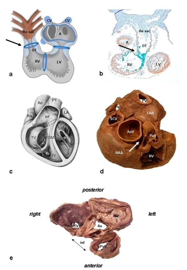
    1. Gittenberger-de Groot AC, et al. Basics of cardiac development for the understanding of congenital heart malformations. Pediatr Res. 2005;57:169. - PubMed
    1. Bartelings MM, et al. Morphogenetic considerations on congenital malformations of the outflow tract. Part 1: Common arterial trunk and tetralogy of Fallot. Int J Cardiol. 1991;32:213. - PubMed
    1. Cai CL, et al. Isl1 identifies a cardiac progenitor population that proliferates prior to differentiation and contributes a majority of cells to the heart. Dev Cell. 2003;5:877. - PMC - PubMed

LinkOut - more resources