

Is it worthwhile to fully evaluate the stomach in every ultrasound examination of the abdominal cavity?
- PMID: 23329910
- PMCID: PMC3522413
Is it worthwhile to fully evaluate the stomach in every ultrasound examination of the abdominal cavity?
Abstract
Background/objective: To evaluate the usefulness of abdominal sonography in the fasting state with no hypotonic agents in the detection and exclusion of gastric lesions.
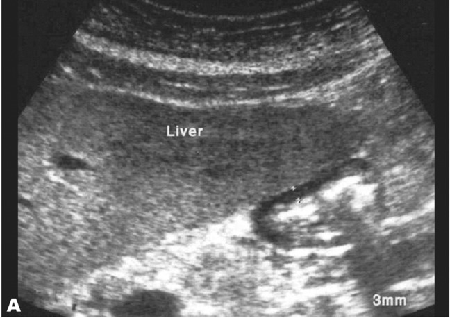
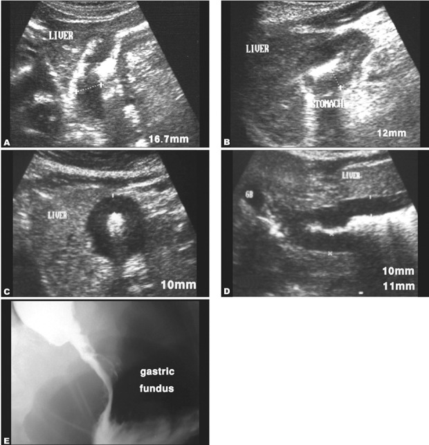
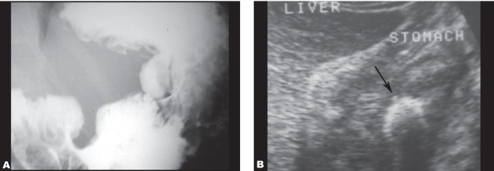
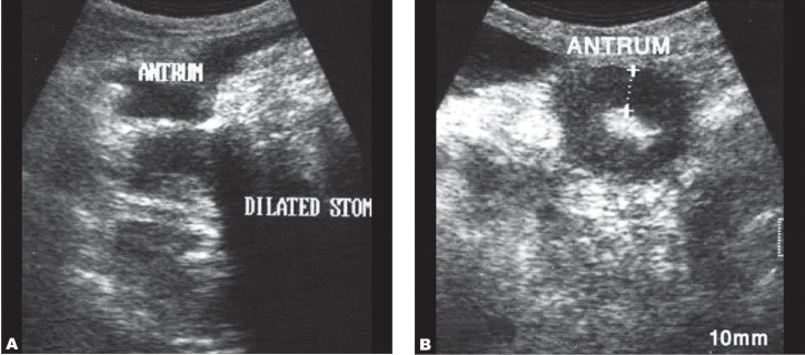
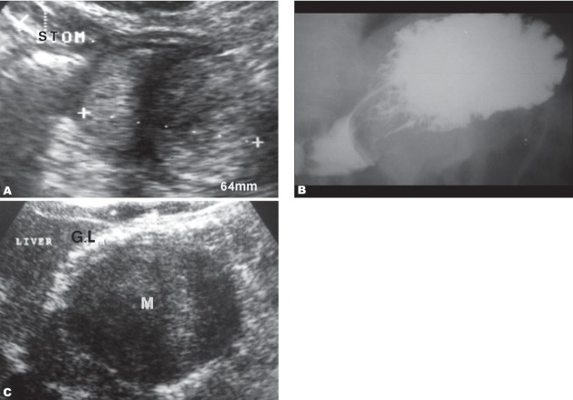
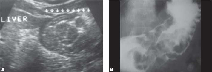
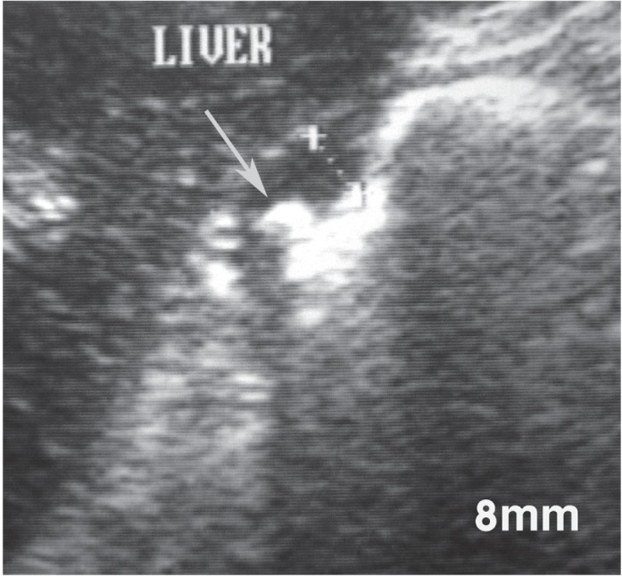
Patients and methods: One-hundred patients with normal upper gastrointestinal endoscopy, 94 patients with a major gastric abnormality (including 59 intraluminal tumors, three submucosal masses, 29 ulcers, two polyps and one hypertrophied gastric mucosa) and 75 patients with minor gastric abnormalities (mainly gastritis) were enrolled into the study.
Results: Of the 100 normal patients, ultrasound showed four false positive results with 96% specificity of the examination. Within the major gastric lesion group, ultrasound was true positive in 55 of 59 tumors, 15 of 29 ulcers, three of three submucosal masses and the case of giant gastric mucosa. It was negative in the detection of gastric polyps. It could detect only 8% of minor gastric abnormalities.
Conclusion: Abdominal sonography in the fasting state, if carefully performed, is sufficiently accurate in detection and exclusion of major gastric lesions. Therefore, although it cannot replace endoscopic and barium studies of the stomach, careful evaluation of the stomach is recommended in every sonographic evaluation of the abdominal cavity.
Keywords: Abdominal Cavity; Gastrointestinal Endoscopy; Sensitivity; Specificity; Ultrasonography.
Figures
References
-
- Lim JH, Lee DH, Ko YT. Sonographic detection of duodenal ulcer. J Ultrasound Med. 1992;11(3):91–4. - PubMed
-
- Fuchs CS, Mayer RJ. Gastric carcinoma. N Engl J Med. 1995 Jul 6;333(1):32–41. - PubMed
-
- Lu CL, Chang SS, Wang SS, Chang FY, Lee SD. Silent peptic ulcer disease: frequency, factors leading to silence, and implications regarding the pathogenesis of visceral symptoms. Gastrointest Endosc. 2004 Jul;60(1):34–8. - PubMed
-
- Tuncel E. Ultrasonic features of duodenal ulcer. Gastrointest Radiol. 1990;15(3):207–10. - PubMed
-
- Puylaert JBCM, van der Zant FM, Rijke AM. Sonography and the acute abdomen: Practical considerations. AJR Am J Roengenol. 1997 Jan;168(1):179–86. - PubMed
LinkOut - more resources
Full Text Sources