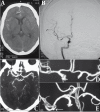
Diagnostic accuracy of early computed tomographic angiography for visualizing medium sized inferior and posterior projecting carotid system aneurysms
- PMID: 23329930
- PMCID: PMC3522323
- DOI: 10.5812/kmp.iranjradiol.17351065.3135
Diagnostic accuracy of early computed tomographic angiography for visualizing medium sized inferior and posterior projecting carotid system aneurysms
Abstract
Background: Conventional angiography, generally referred to as intra-arterial digital subtraction angiography, still remains the gold standard reference method for the diagnosis of intracranial aneurysms, helical computed tomography angiography (CTA) is a new non-invasive volumetric imaging method.
Objectives: This study was conducted to screen patients presenting with subarachnoidhemorrhage by CTA before conventional digital subtraction angiography (DSA) and subsequently comparing the results for various aneurysm projections.
Patients and methods: In a prospective study, 99 consecutive patients with an initial diagnosis of subarachnoid hemorrhage were screened for aneurysms with CTA followed by conventional DSA. There were 17 cases with negative angiograms in whom repeat angiograms, three months later were negative for 15 cases, while two cases were found to bear aneurysm on the repeat examination. Eighty two patients had at least one proven aneurysm on initial DSA and two on the repeat angiogram. Out of 84 patients, five underwent endovascular treatment and 79 patients who underwent surgical clipping were considered for projection evaluation.
Results: Sensitivity of CTA was 98.78% (95% confidence interval [CI], 93.4-99.7%), while the specificity was 100% (95% CI, 81.57-100%) and the kappa coefficient of agreement between CTA and DSA was 96.5%. The most significant discrepancies with DSA findings were for visualizing the projection of inferior and posterior projecting proximal anterior circulation aneurysms.
Conclusions: Helical CTA was in good concordance with DSA for screening of cerebral aneurysms; however, for exact visualization of the aneurysm neck and its projection, especially if it is inferior or posterior, DSA remains the gold standard.
Keywords: Angiography; Digital Subtraction; Helical CT; Intracranial Aneurysm.
Figures

References
-
- King JT, Jr. Epidemiology of aneurysmal subarachnoid hemorrhage. Neuroimaging Clin N Am. 1997;7(4):659–68. - PubMed
-
- Korogi Y, Takahashi M, Mabuchi N, Nakagawa T, Fujiwara S, Horikawa Y, et al. Intracranial aneurysms: diagnostic accuracy of MR angiography with evaluation of maximum intensity projection and source images. Radiology. 1996;199(1):199–207. - PubMed
LinkOut - more resources
Full Text Sources
