Case Reports
. 2011 Nov;8(3):182-9.
doi: 10.5812/kmp.iranjradiol.17351065.3392. Epub 2011 Nov 25.

Radiological features of osteoid osteoma: pictorial review

Affiliations
Case Reports

Radiological features of osteoid osteoma: pictorial review

Jahanbakhsh Hashemi et al. Iran J Radiol. 2011 Nov.

Abstract

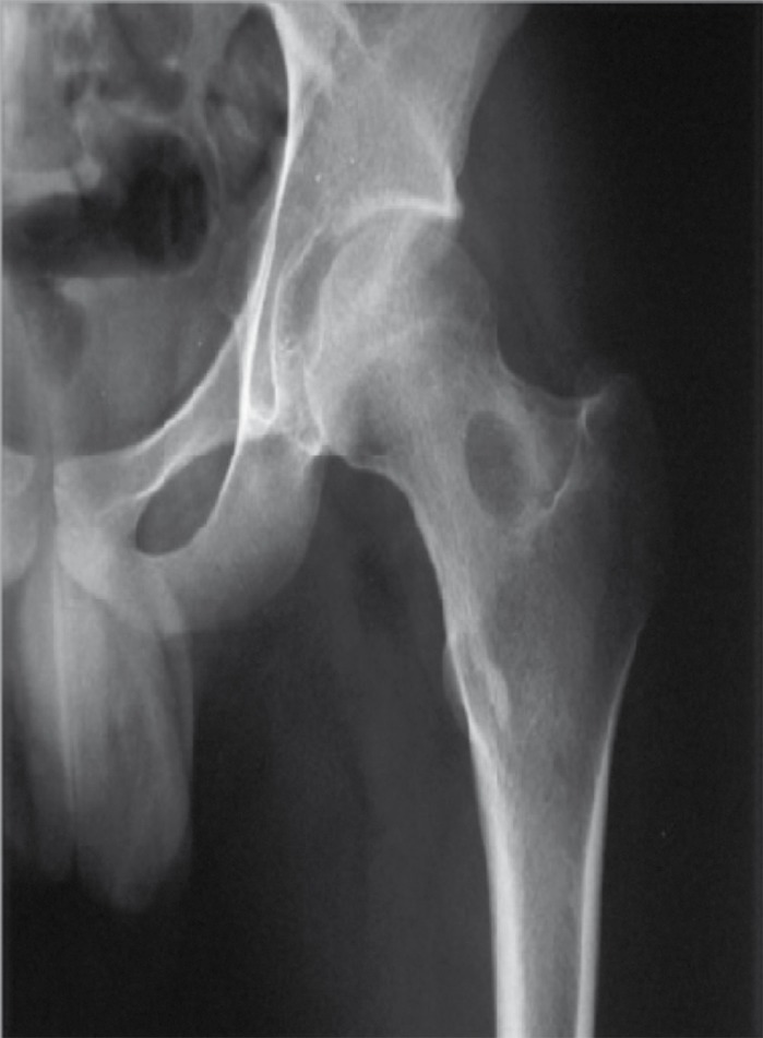
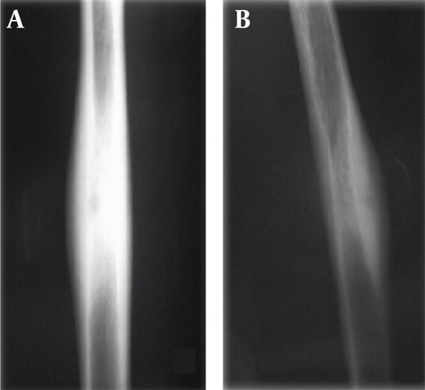
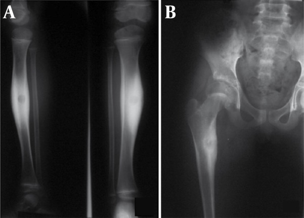
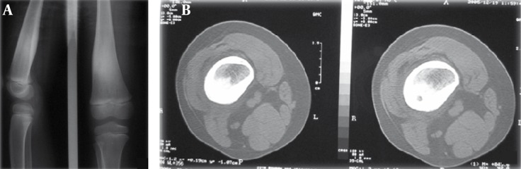
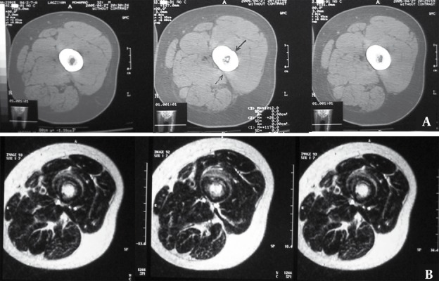
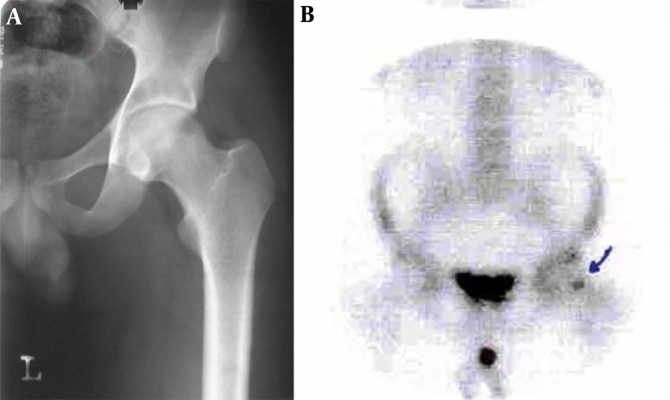
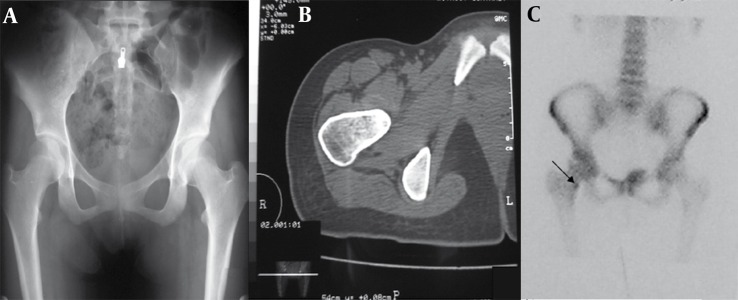
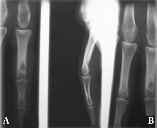
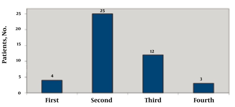
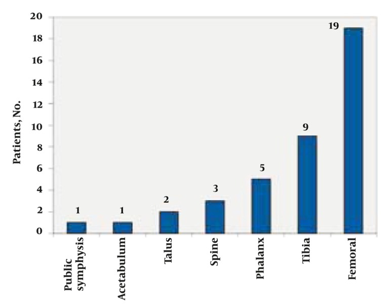
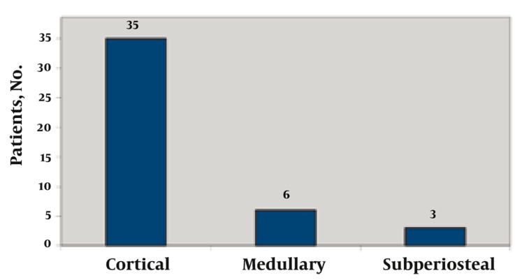
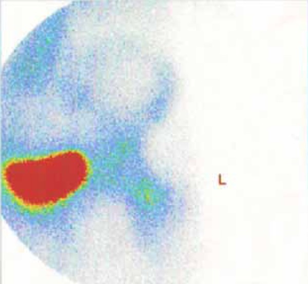
Osteoid osteoma is a benign bone tumor of undetermined etiology, composed of a central zone named nidus which is an atypical bone completely enclosed within a wellvascularized stroma and a peripheral sclerotic reaction zone. There are three types of radiographic features: cortical, medullary and subperiosteal. Forty-four patients with osteoid osteoma were studied retrospectively. In plain films, 35 patients presented as the cortical type, six cases were located in the medullary zone and three had subperiosteal osteoid osteoma. In all the cases, the nidus was visualized on computed tomography (CT) scan. The nidus was visible in four out of five patients who had also undergone magnetic resonance imaging (MRI). Double-density sign, seen on radionuclide bone scans was positive in all patients. MRI is more sensitive in the diagnosis of bone marrow and soft tissue abnormalities adjacent to the lesion, and in the nidus that is located closer to the medullary zone. On the other hand, CT is more specific when it comes to detecting the lesion's nidus.

Keywords: Magnetic Resonance Imaging; Osteoma, Osteoid; Radionuclide Imaging; Tomography, X-Ray Computed.


Figures

Figure 1
Figure 1. A 19-Year-Old Man With Pain in the Thigh.
Body section film reveals a circular area of density with a central radiolucent nidus in the femoral neck.
Figure 2
Figure 2. A 17-Year-Old Man With Pain in His Arm.
The lesion is characterized as a small nidus surrounded by sclerotic bone. A, The nidus is not demonstrated in the AP projection but B, Precisely in the lateral projection
Figure 3
Figure 3. A, A 14-Year-Old Man With Leg Pain; and B, An 18-Year-Old Female With Pain in Her Thigh.
In the plain film the nidus is calcified and surrounded by increased bone density.
Figure 4
Figure 4. A 17-Year-Old Man Presenting With Thigh Pain.
A, Plain radiographs show endosteal thickening at the distal of the femur. No definite radiolucencies are present. B, CT scan reveals the endosteal thickening and demonstrates radiolucencies in the cortex.
Figure 5
Figure 5. A 25-Year-Old Man Complaining of Pain in Mid FemurA.
A, CT scan through mid-femur. Nidus is visible in the medulary region only in one of the cuts. B, MRI of the same patient reveals distinct hypersignal nidus in the medulary region in T2-W sequence in several cuts.
Figure 6
Figure 6. A 28-Year-Old Man Presenting With Thigh Pain.
A, Plain radiograghs show no pathologic finding in cortex and medulla such as nidus or peripheral sclerotic reaction. B, Technetium scan demonstrates increased uptake in the area of the nidus (arrow). Biopsy of the mentioned lesion confirmed the diagnosis of osteoid osteoma.
Figure 7
Figure 7. A 20-Year-Old Woman Presented With a One-Year Right Hip Pain.
A, Plain radiograph of the hip revealed no abnormalities. B, CT scan of the hip shows a sclerotic lesion with a central nidus in the cortex. C, In radionuclide scan an increased uptake was detected in the same region (arrow).
Figure 8
Figure 8. A 28-Year-Old Man With Pain in the Hand.
A, Plain radiograghs show osteoid osteoma of the distal of the phalanx (medullary type) in AP and B, Lateral projection.
Figure 9
Figure 9. A 17-Year-Old Man Presenting With Foot Pain.
Plain film shows subperiosteal osteoid osteoma of the talus which often presents as a round soft tissue mass adjacent to bone.
Figure 10
Figure 10. A 22-Year-Old Man With a Complaint of 5-Month Continuous Low Back Pain
A, Radiograph revealed a sclerotic region in left pedicle of L1 vertebra along with scoliosis in thoracolumbar region. B, Lumbar CT scan revealed osteolytic and osteoblastic lesion in pedicle of L1 vertebra. C, MRI with T1 weighting demonstrated focal low signal intensity surrounded by soft tissue component replaced pedicle of L1 vertebra on the left side in the axial sections. Diffuse low signal intensity due to edema was seen in the adjacent bone.
Figure 11
Figure 11. A Nine-Year-Old Boy With the Complaint of Right Shin Pain.
A, The AP and lateral radiograph of the right shin revealed a solid periosteal reaction in the medial part of tibia without any obvious nidus. B, CT scan of the mid part of the right shin revealed a nidus along with a periosteal reaction around the lesion.
Figure 12
Figure 12. Distribution of Patients Based on Age Decade at Presentation
Figure 13
Figure 13. Osteoid Osteoma, Anatomical Sites of Origin Among 44 Patients
Figure 14
Figure 14. Distribution of Disease According to the Site of Origin
Figure 15
Figure 15. Double-Density Sign in the Femoral Neck

References

    1. Edeiken J, Dalinka MK, Karasick D. Edeiken’s roentgen diagnosis of diseases of bone. Baltimore: Williams & Wilkins; 1990.
    1. Helms CA, Hattner RS, Vogler JB, 3rd. Osteoid osteoma: radionuclide diagnosis. Radiology. 1984;151(3):779–84. - PubMed
    1. O’Connell JX, Nanthakumar SS, Nielsen GP, Rosenberg AE. Osteoid osteoma: the uniquely innervated bone tumor. Mod Pathol. 1998;11(2):175–80. - PubMed
    1. Gaeta M, Minutoli F, Pandolfo I, Vinci S, D’Andrea L, Blandino A. Magnetic resonance imaging findings of osteoid osteoma of the proximal femur. Eur Radiol. 2004;14(9):1582–9. doi: 10.1007/s00330-004-2293-5. - DOI - PubMed
    1. Allen SD, Saifuddin A. Imaging of intra-articular osteoid osteoma. Clin Radiol. 2003;58(11):845–52. doi: 10.1016/S0009-9260(03)00213-7. - DOI - PubMed

Publication types

LinkOut - more resources