. 2013;6(2):124-41.
Epub 2013 Jan 15.

Matrix metalloproteinase inhibition negatively affects muscle stem cell behavior

Affiliations

Matrix metalloproteinase inhibition negatively affects muscle stem cell behavior

Ian Bellayr et al. Int J Clin Exp Pathol. 2013.

Abstract

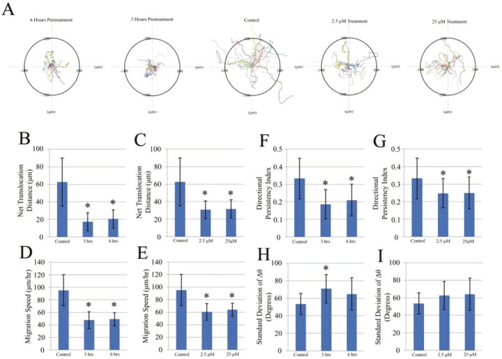
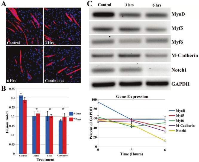
Skeletal muscle is a large and complex system that is crucial for structural support, movement and function. When injured, the repair of skeletal muscle undergoes three phases: inflammation and degeneration, regeneration and fibrosis formation in severe injuries. During fibrosis formation, muscle healing is impaired because of the accumulation of excess collagen. A group of zinc-dependent endopeptidases that have been found to aid in the repair of skeletal muscle are matrix metalloproteinases (MMPs). MMPs are able to assist in tissue remodeling through the regulation of extracellular matrix (ECM) components, as well as contributing to cell migration, proliferation, differentiation and angiogenesis. In the present study, the effect of GM6001, a broad-spectrum MMP inhibitor, on muscle-derived stem cells (MDSCs) is investigated. We find that MMP inhibition negatively impacts skeletal muscle healing by impairing MDSCs in migratory and multiple differentiation abilities. These results indicate that MMP signaling plays an essential role in the wound healing of muscle tissue because their inhibition is detrimental to stem cells residing in skeletal muscle.

Keywords: MMPs; Skeletal muscle stem cells; cell migration; differentiation.


Figures

Figure 1
Figure 1
Cell migration is reduced with the administration of GM6001. A-I: phase contrast images taken of MDSCs using a live automated cell imager at 1 (A, D, G), 3 (B, E, H) and 6 (C, F, I) hours of MDSCs pretreated for 3 hours with 25μM GM6001 prior to an artificial wound (A, B, C), control group with no treatment (D, E, F), and 25μM GM6001 added to cell culture after artificial wound (G, H, I). The red line indicates the initial edge of the wound and the green line indicates the position of the cells after 1, 3 or 6 hours of migration into the wound area. Statistical analysis of MDSC (J, K) and C2C12 myoblast (L, M) migration based on pretreatment time with 25μM GM6001 and concentration (0, 2.5, 25μM) after the creation of an artificial wound. There is a significant difference (*P<0.05) from the non-treated group (control) at each time point.
Figure 2
Figure 2
Tracking a single cell migration with GM6001 treatment. The migration paths of 20 individual MDSCs of different experimental groups captured in a time-lapse motility assay (A, data was pooled from three independent experiments). The net translocation distance (straight distance from the start to the end point) of each single MDSC over a 2 hour period is represented as the mean ± standard deviation of the paths of 20 randomly selected cells that were either pretreated with 25 μM of GM6001 prior to image capture (B) or treated with different concentrations at the start of image capture (C). The migration speed (total length of the migration path per hour) of each cell is shown as the mean ± standard deviation of 20 randomly selected cells that were either pretreated with 25 μM of GM6001 prior to image capture (D) or treated with different concentrations at the start of image capture (E). The directional persistency index (ratio of the net translocation distance to the cumulative length of the migration path) of each MDSC over a 2 hour period is represented at the mean ± standard deviation of the paths of 20 randomly selected cells that were either pretreated with 25 μM of GM6001 prior to image capture (F) or treated with different concentrations at the start of image capture (G). The centroid directional movement (a measure of the change in the direction of the centroid movement of a single cell) is shown as the mean ± standard deviation of 20 randomly selected cells that were either pretreated with 25 μM of GM6001 prior to image capture (H) or treated with different concentrations at the start of image capture (I). There is a significant difference (*P<0.05) from the non-treated group (control) at each time point.
Figure 3
Figure 3
GM6001 has no effect to muscle cell proliferation. The population doubling time of MDSCs and C2C12 myoblasts were unaffected based on pretreatment time (A) and different concentrations (B) of GM6001 administered to the cells. C2C12 cells (D) exhibit a greater expression of EdU (red) compared to MDSCs (C) regardless of MMP inhibition pretreatment (E) or different concentrations (F). All cells were stained with Hoechst 33342 (blue).
Figure 4
Figure 4
Stem cell characterizes’ change with GM6001 treatment. The gene expression of MDSCs treated with 25μM of GM6001 for 3 and 6 hours. These results indicate that MDSCs treated with an MMP inhibitor had reduced expression of stem cell markers, CD34 and Sca1. GAPDH was used as a loading control.
Figure 5
Figure 5
GM6001 interacts muscle stem cells’ multiple differentiations. MDSC treated with GM6001 exhibited a reduced osteogenic differentiation potential based on staining for ALP, Von Kossa and Alizarin Red. The white arrows denote calcium deposition for Von Kossa stains and black arrows denote the red from Alizarin red stains. MDSCs treated with GM6001 (2.5μM and 25μM) exhibited reduced accumulation of lipids (red) within the cytoplasm after 2 weeks as shown with Oil Red O staining. The percentage of ALP positive MDSCs after 3 days of osteogenic differentiation is reduced with MMP inhibition (B). The percentage of MDSCs positive for EdU expression during osteogenic differentiation (C). The optical density was used to quantify adipogenesis (D). There is a significant difference (*P<0.05) between the control and other groups. Statistical significant between control and treated groups was determined if *P<0.05.
Figure 6
Figure 6
GM6001 inhibits myogenic differentiation in vitro. MMP inhibitor, GM6001, reduces muscle cell myotube formation (A). Immunostaining of MHC (red) in MDSC. There is a significant difference (*P<0.05) between GM6001 treated MDSCs and the control at each time point (B). MMP inhibition negatively impacts genes associated with myotube formation in MDSCs (C). Gene expression was examined for 4 genes related to myogenic differentiation (MyoD, Myf5, Myf6 and M-cadherin) and 1 signaling gene (Notch1) after 3 and 6 hours pretreatment with GM6001 and 1 day cultured with myogenic differentiation meda. GAPDH was used as loading control.
Figure 7
Figure 7
GM6001 inhibits muscle repair processes in vivo. Pax7 expression (red) was reduced 5 days after laceration injury with GM6001 administration in comparison to non-treated laceration injuries (A). Dystrophin (green) and cell nuclei (blue) were also counterstained. GM6001 treatment of injured mouse skeletal muscle tissue exhibits elevated levels of collagen deposition 7 days post laceration injury than the non-treated skeletal muscle tissue (B). Masson’s modified trichrome staining was used to identify normal healthy skeletal muscle tissue (red) and collagen (blue). While an elevated level of collagen deposition was observed 7 and 12 days after laceration injury, there was no significant difference (C).

Similar articles

Cited by

References

    1. Charge SB, Rudnicki MA. Cellular and molecular regulation of muscle regeneration. Physiol Rev. 2004;84:209–38. - PubMed
    1. Grefte S, Kuijpers-Jagtman AM, Torensma R, Von den Hoff JW. Skeletal muscle development and regeneration. Stem Cells Dev. 2007;16:857–68. - PubMed
    1. Huard J, Li Y, Fu FH. Muscle injuries and repair: current trends in research. J Bone Joint Surg Am. 2002;84-A:822–32. - PubMed
    1. Nozaki M, Li Y, Zhu J, Ambrosio F, Uehara K, Fu FH, Huard J. Improved muscle healing after contusion injury by the inhibitory effect of suramin on myostatin, a negative regulator of muscle growth. Am J Sports Med. 2008;36:2354–62. - PubMed
    1. Ritenour AE, Christy RJ, Roe JL, Baer DG, Dubick MA, Wade CE, Holcomb JB, Walters TJ. The Effect of a Hypobaric, Hypoxic Environment on Acute Skeletal Muscle Edema after Ischemia-Reperfusion Injury in Rats. Journal of Surgical Research. 2010;160:253–259. - PubMed

Publication types

MeSH terms

Substances

LinkOut - more resources