

A clinicopathologic study of esophageal 860 benign and malignant lesions in 910 cases of consecutive esophageal biopsies
- PMID: 23330004
- PMCID: PMC3544238
A clinicopathologic study of esophageal 860 benign and malignant lesions in 910 cases of consecutive esophageal biopsies
Abstract
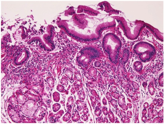
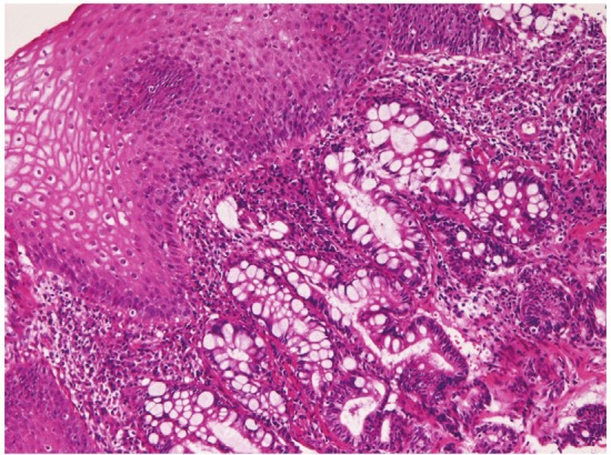
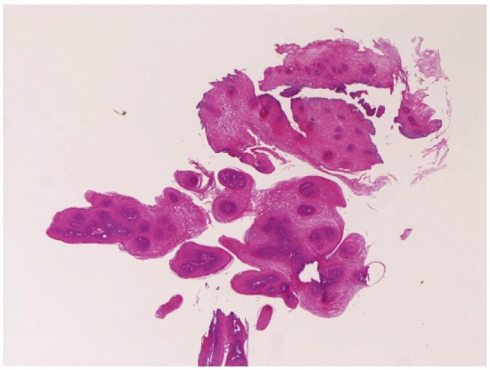
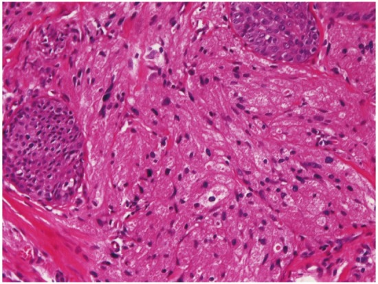
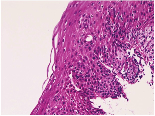
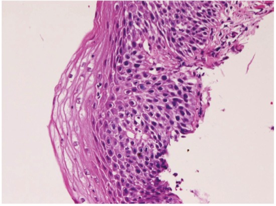
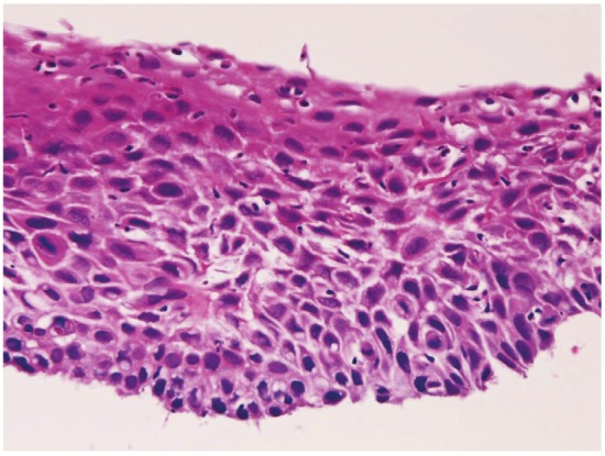
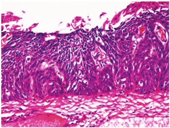
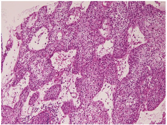
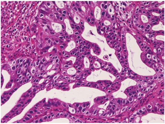
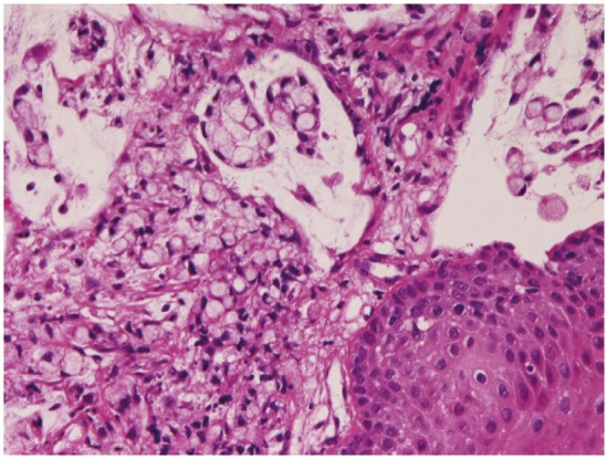
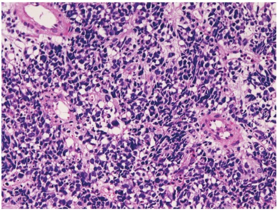
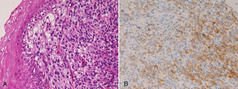
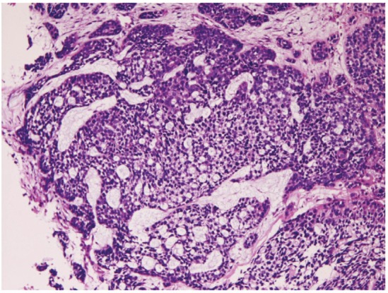
The author reviewed 910 cases of consecutive esophageal biopsies in the last 15 year in the pathology laboratory of our hospital. There were 693 normal mucosa and benign lesions (76.2%) and 217 malignant lesions (23.8%). No significant changes were recognized in the esophagus in 50 biopsies (5.5%). In benign lesions, the number and frequency (percentages) were as follows: 263 chronic esophagitis (28.9%), 98 heterotopic gastric mucosa (10.8%), 3 heterotopic colonic mucosa (0.3%), 71 glycogenic acanthosis (7.8%), 68 candidiasis (7.5%), 35 benign ulcer (3.8%), 41 squamous papilloma (4.5%), 4 granular cell tumor (0.4%), 1 tubular adenoma (0.1%), 2 cytomegalovirus esophagitis (0.2%), 3 leiomyoma (0.3%), 17 basal cell hyperplasia (1.9%), and 37 Barrett's epithelium (4%). In malignant lesions, the number and frequency (percentages) were as follows: 53 mild dysplasia (5.8%), 29 moderate dysplasia (3.2%), 31 severe dysplasia (3.4%), 13 carcinoma in situ (1.4%), 68 squamous cell carcinoma (7.5%), 7 primary adenocarcinoma (0.8%), 1 primary signet ring cell carcinoma (0.1%), 4 primary small cell carcinoma (0.4%), 2 primary amelanotic malignant melanoma (0.2%), 1 primary undifferentiated sarcoma (0.1%), 7 gastric cancer invasion (0.8%), and 1 primary adenoid cystic carcinoma (0.1%). In this article, the clinicopathologic features of these esophageal lesions were described.
Keywords: Esophagus; benign lesions; clinicopathologies; immunohistochemistry; malignant lesions.
Figures
References
-
- Rosai J. Rosai and Ackermann’s Pathology. Ninth edition. Mosby; Esophagus; pp. 615–647.
-
- Hamilton SR, Aaltonen LA, editors. WHO Classification of tumors. Pathology and genetics, Tumor of the digestive system. Lyon: IARC press; 2000. Chapter 1, tumors of oesophagus; pp. 10–30.
-
- Terada T, Kawaguchi M. Primary clear cell adenocarcinoma of the peritoneum. Tohoku J Exp Med. 2005;206:271–275. - PubMed
-
- Terada T, Kawaguchi M, Furukawa K, Sekido Y, Osamura Y. Minute mixed ductal-endocrine carcinoma of the pancreas with predominant intraductal growth. Pathol Int. 2002;52:740–746. - PubMed
-
- Terada T, Tanigichi M. Intraductal oncocytic papillary neoplasm of the liver. Pathol Int. 2004;54:116–123. - PubMed
MeSH terms
LinkOut - more resources
Full Text Sources
Medical