. 2013 Feb 19;85(4):2208-15.
doi: 10.1021/ac3029344. Epub 2013 Feb 5.

Elastomeric negative acoustic contrast particles for affinity capture assays

Affiliations

Elastomeric negative acoustic contrast particles for affinity capture assays

Kevin W Cushing et al. Anal Chem. .

Abstract

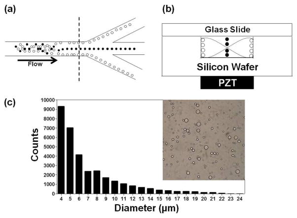
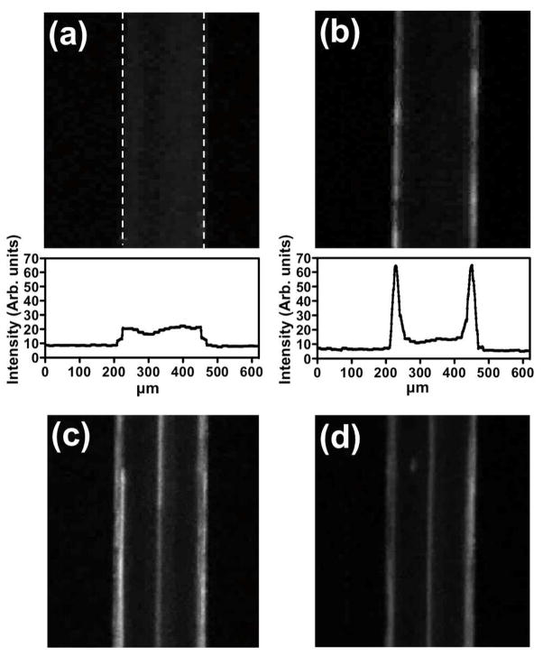
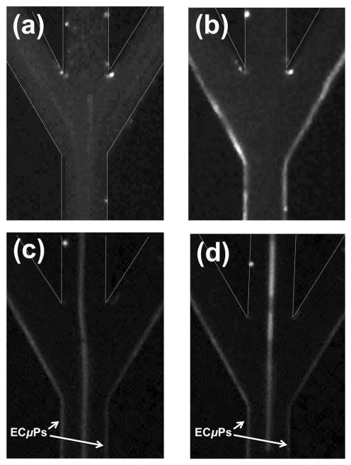
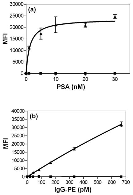
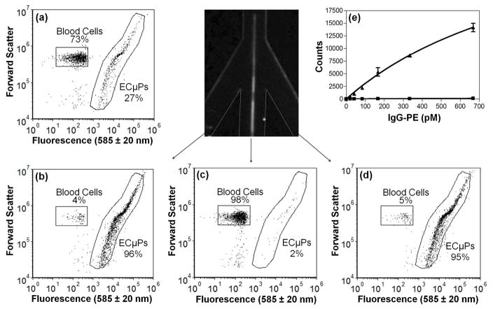
This report describes the development of elastomeric capture microparticles (ECμPs) and their use with acoustophoretic separation to perform microparticle assays via flow cytometry.We have developed simple methods to form ECμPs by cross-linking droplets of common commercially available silicone precursors in suspension followed by surface functionalization with biomolecular recognition reagents. The ECμPs are compressible particles that exhibit negative acoustic contrast in ultrasound when suspended in aqueous media, blood serum, or diluted blood. In this study, these particles have been functionalized with antibodies to bind prostate specific antigen and immunoglobulin (IgG). Specific separation of the ECμPs from blood cells is achieved by flowing them through a microfluidic acoustophoretic device that uses an ultrasonic standing wave to align the blood cells, which exhibit positive acoustic contrast, at a node in the acoustic pressure distribution while aligning the negative acoustic contrast ECμPs at the antinodes. Laminar flow of the separated particles to downstream collection ports allows for collection of the separated negative contrast (ECμPs) and positive contrast particles (cells). Separated ECμPs were analyzed via flow cytometry to demonstrate nanomolar detection for prostate specific antigen in aqueous buffer and picomolar detection for IgG in plasma and diluted blood samples. This approach has potential applications in the development of rapid assays that detect the presence of low concentrations of biomarkers in a number of biological sample types.


Figures

Figure 1
Figure 1
(a) Schematic diagram depicting the separation approach for elastomeric negative acoustic contrast particles (white) from positive acoustic contrast particles (e.g., blood cells) (black) at the trifurcation in a silicon acoustic sample preparation chip. (b) Cross-section, at dashed line in (a), of the chip, with the positive contrast particles focused at the pressure node and the negative contrast particles focused at the pressure antinodes under an acoustic standing wave field. Note: Images are not drawn to scale. (c) Bright field image and size histogram of PDMS-based elastomeric particles prepared by bulk emulsification.
Figure 2
Figure 2
ECμPs function as negative contrast particles and can be separated from positive contrast particles using an acoustic sample preparation chip. These images were captured via an epifluorescence microscope with a 2.5x objective. Each sample was flowing at 45 μL/min. Fluorescence microscopy images along with histograms of average intensity profiles for Nile Red (NR) stained ECμPs with the field (a) off and then (b) on. (c) NR-ECμPs separated from NR-PS particles with the field on. (d) NR-ECμPs separated from NR-blood cells with the field on. Note: white dashed lines in (a) indicate micro-channel borders.
Figure 3
Figure 3
(a) Fluorescence microscopy images of NR-ECμPs with the field (a) off and then (b) on. (c) NR-ECμPs separated from NR-PS particles with the field on. (d) NR-ECμPs separated from NR-blood cells (0.1% volume whole porcine blood) with the field on. Each sample was flowing at 45 μL/min. Note: white dashed lines indicate micro-channel borders that are not otherwise visible due to presence of NR-ECμPs.
Figure 4
Figure 4
ECμPs as platforms for protein capture assays in flow cytometry. Note: (▲)denotes ECμPs with capture antibody and (■) denotes particles without capture antibody. (a) ECμPs used in a sandwich assay for prostate specific antigen (PSA) in physiological buffer (PBS). (b) ECμPs used in a binding assay for goat anti-mouse IgG-phycoerythrin performed in 10% volume porcine plasma diluted with PBS. Note: all data points were obtained from triplicate experiments analyzed by gated flow cytometric analysis. Error bars represent the standard deviation of the mean for 3 separate determinations of median fluorescence intensity.
Figure 5
Figure 5
ECμPs used in an assay in diluted blood where acoustic separation and collection was achieved using the acoustic sample preparation chip prior to flow cytometry analysis. (a)Flow cytometry scatter plot (forward scatter versus fluorescence (585 ± 20 nm)) showing the initial mixture (inlet) of ligand-bound ECμPs and blood cells. (b) Scatter plot of the collected fraction of the left peripheral outlet channel. (c) Scatter plot of the collected fraction from the central outlet channel. (d) Scatter plot of the collected fraction of the right peripheral outlet channel. (e) IgG-PE binding assay in 0.1 % porcine blood from ECμPs separated and collected using an acoustic sample preparation chip, prior to flow cytometry analysis. (▲) denotes ECμPs with capture antibody and (■) denotes particles without capture antibody. Note: all data points were obtained in triplicate. Error bars represent the standard deviation of the mean for 3 separate determinations of median fluorescence intensity.

References

    1. Laurell T, Petersson F, Nilsson A. Chip integrated strategies for acoustic separation and manipulation of cells and particles. Chemical Society Reviews. 2007;36(3):492–506. - PubMed
    1. Nam J, Lim H, Kim D, Shin S. Separation of platelets from whole blood using standing surface acoustic waves in a microchannel. Lab on a Chip. 2011;11(19):3361–3364. - PubMed
    1. Gossett DR, Weaver WM, Mach AJ, Hur SC, Tse HTK, Lee W, Amini H, DiCarlo D. Label-free cell separation and sorting in microfluidic systems. Analytical and bioanalytical chemistry. 2010;397(8):3249–3267. - PMC - PubMed
    1. Piyasena ME, Suthanthiraraj PPA, Applegate RW, Jr, Goumas AM, Woods TA, Lopez GP, Graves SW. Multinode Acoustic Focusing for Parallel Flow Cytometry. Analytical Chemistry. 2012;84(4):1831–1839. - PMC - PubMed
    1. Meng Z, Veenstra TD. Targeted mass spectrometry approaches for protein biomarker verification. Journal of Proteomics. 2011;74(12):2650–2659. - PubMed

Publication types