. 2013 Jan 18:9:15.
doi: 10.1186/1746-6148-9-15.

The effects of taurolidine alone and in combination with doxorubicin or carboplatin in canine osteosarcoma in vitro

Affiliations

The effects of taurolidine alone and in combination with doxorubicin or carboplatin in canine osteosarcoma in vitro

Kevin Marley et al. BMC Vet Res. .

Abstract

Background: Osteosarcoma (OS) affects over 8000 dogs/year in the United States. The disease usually arises in the appendicular skeleton and metastasizes to the lung. Dogs with localized appendicular disease benefit from limb amputation and chemotherapy but most die within 6-12 months despite these treatments. Taurolidine, a derivative of taurine, has anti-tumor and anti-angiogenic effects against a variety of cancers. The following in vitro studies tested taurolidine as a candidate for adjuvant therapy for canine OS. Tests for p53 protein status and caspase activity were used to elucidate mechanisms of taurolidine-induced cell death.

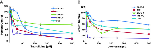
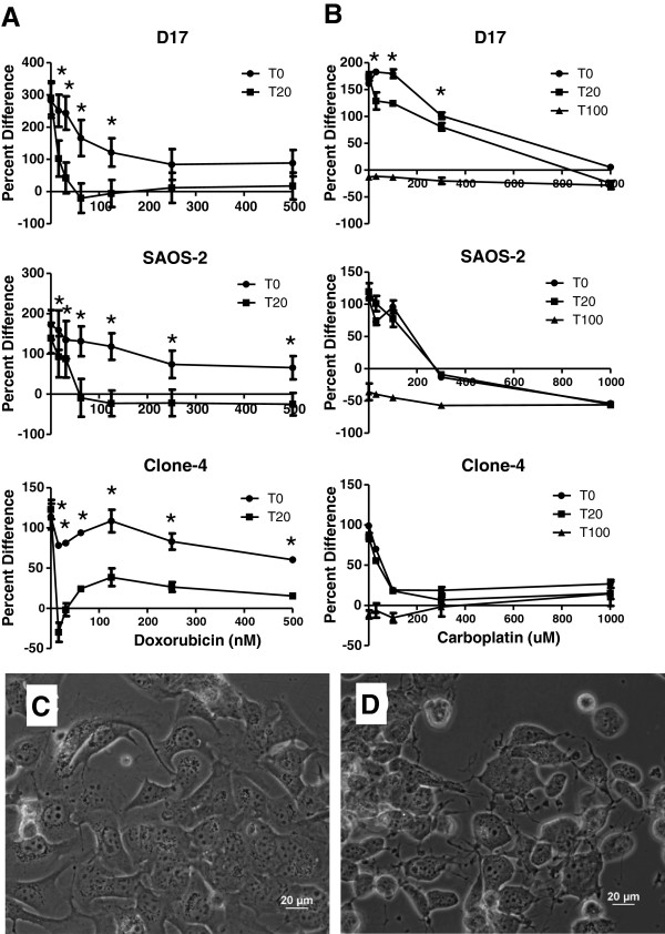
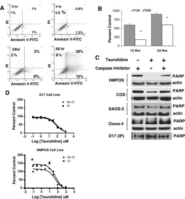
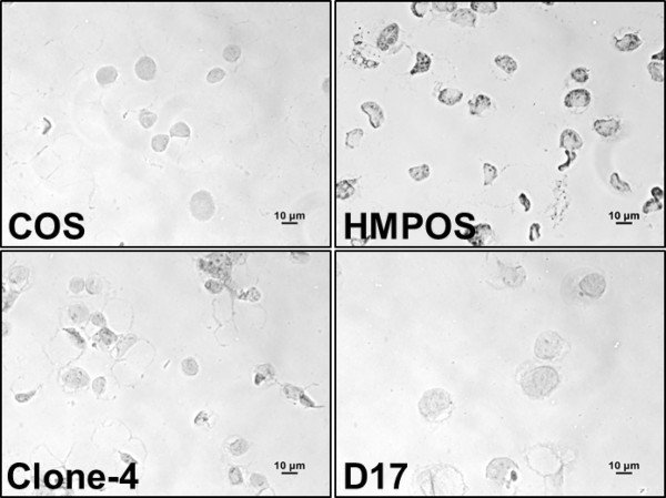
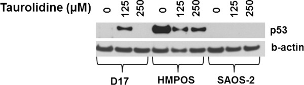
Results: Taurolidine was cytotoxic to osteosarcoma cells and increased the toxicity of doxorubicin and carboplatin in vitro. Apoptosis was greatly induced in cells exposed to 125 μM taurolidine and less so in cells exposed to 250 μM taurolidine. Taurolidine cytotoxicity appeared caspase-dependent in one cell line; with apparent mutant p53 protein. This cell line was the most sensitive to single agent taurolidine treatment and had a taurolidine-dependent reduction in accumulated p53 protein suggesting taurolidine's effects may depend on the functional status of p53 in canine OS.

Conclusion: Taurolidine's cytotoxic effect appears dependent on cell specific factors which may be explained, in part, by the functional status of p53. Taurolidine initiates apoptosis in canine OS cells and this occurs to a greater extent at lower concentrations. Mechanisms of cell death induced by higher concentrations were not elucidated here. Taurolidine combined with doxorubicin or carboplatin can increase the toxicity of these chemotherapy drugs and warrants further investigation in dogs with osteosarcoma.


Figures

Figure 1
Figure 1
Effects of taurolidine or doxorubicin on proliferation of OS cell lines. Cells were incubated for 24 hrs with taurolidine A) or 72 hrs with doxorubicin B) and cytotoxic effects were determined using an MTS assay. Error bars indicate SD of 4 replicate wells. In some cases error bars are too small to be visible.
Figure 2
Figure 2
Effects of doxorubicin or carboplatin ± taurolidine on proliferation of OS cell lines. Three OS cell lines were incubated for 72 hrs in increasing concentrations of doxorubicin A) or carboplatin B) in the presence or absence of taurolidine and cytotoxic effects were evaluated using an MTS assay. T0 indicates no taurolidine while T20 and T100 indicate 20 μM and 100 μM taurolidine respectively. Error bars indicate SD of 4 replicate wells. In some cases error bars are too small to be visible. * indicates a synergistic interaction (see methods). Data are presented as percent difference from time 0 on the y-axis (see methods and results). Photomicrographs of D17 cells after 6 hr incubation with 100 nM doxorubicin C) or with 100 nM doxorubicin plus 20 μM taurolidine. D) Cytoplasmic contraction is readily appreciated in the cells exposed to the drug combination and represents an early apoptotic effect.
Figure 3
Figure 3
Mechanisms of taurolidine-induced cytotoxicity are cell line and concentration dependent. Binding of annexin-V-biotin-FITC and propidium iodide (PI) to taurolidine-treated D17 OS cells was determined by 2-color flow cytometry. A) Data are displayed as dot plots and data points in the lower right quadrant (i.e., annexin-V positive) represent early apoptotic cells while data points in the upper left quadrant represent necrotic cells and points in the upper right quadrant (dual annexin-V/PI positive) represent cells in late apoptosis and necrosis. B) The release of nucleosomes as an indicator of apoptosis following taurolidine treatment of D17 OS cells was determined at two time points. Data are expressed as percent of apoptosis values measured in untreated cells at the same time points. The higher taurolidine concentration (250 μM) was less effective at inducing apoptotic changes in this assay indicating cell death occurred by other mechanisms. T125, T250 = 125 μM and 250 μM taurolidine respectively. * indicates different from 125 μM at same time point (p < .01). Error bars indicate SD. C) Western blots show degradation of PARP as an indirect indicator of taurolidine-activated caspase activity in COS and HMPOS cells. Cells incubated 12 hrs in taurolidine as described in methods. No loading control was available for the D17 blot because it was an immunoprecipitation. D) These proliferation assays show the caspase inhibitor (CI) Z-VAD-FMK protects HMPOS but not D17 OSA cells from caspase-dependent cell death at low concentrations of taurolidine, consistent with PARP cleavage seen in 3C. * indicates intercept is different between treated and untreated conditions (P < .05).
Figure 4
Figure 4
p53 protein accumulation in untreated osteosarcoma cells. Representative photomicrographs of cells probed with an antibody against p53 protein show strong accumulation in HMPOS cells, moderate staining in Clone-4 and none in the D17 and COS cell lines. The signal that can be observed in D17 and COS cells is the counterstain hematoxylin (see methods).
Figure 5
Figure 5
Effects of taurolidine on p53 protein expression. Western blots showing the effects of taurolidine exposure on this cell cycle regulatory protein is concentration and cell line dependent. The taurolidine-induced appearance of p53 protein in D17 cells suggests these cells proceed through a p53-induced apoptotic pathway when exposed to 125 uM taurolidine. In HMPOS cells, with apparent mutant p53, p53 was down-regulated suggesting cell death may occur through different pathways in this cell line. Clone 4 and COS cells did not show any p53 expression at any concentration of taurolidine tested (data not shown).

Similar articles

Cited by

References

    1. Royals SR, Farese JP, Milner RJ, Lee-Ambrose L, van Gilder J. Investigation of the effects of deracoxib and piroxicam on the in vitro viability of osteosarcoma cells from dogs. Am J Vet Res. 2005;66(11):1961–1967. doi: 10.2460/ajvr.2005.66.1961. - DOI - PubMed
    1. Imhof L, Goldinger SM, Baumann K, Schad K, French LE, Rothlisberger P, Dummer R. The antibacterial substance, taurolidine in the second/third-line treatment of very advanced stage IV melanoma including brain metastases: results of a phase 2, open-label study. Melanoma Res. 2011;21(1):80–83. doi: 10.1097/CMR.0b013e328341442d. - DOI - PubMed
    1. Chromik AM, Daigeler A, Bulut D, Flier A, May C, Harati K, Roschinsky J, Sulberg D, Ritter PR, Mittelkotter U. et al.Comparative analysis of cell death induction by Taurolidine in different malignant human cancer cell lines. J Exp Clin Cancer Res. 2010;29:21. doi: 10.1186/1756-9966-29-21. - DOI - PMC - PubMed
    1. Braumann C, Jacobi CA, Rogalla S, Menenakos C, Fuehrer K, Trefzer U, Hofmann M. The tumor suppressive reagent taurolidine inhibits growth of malignant melanoma–a mouse model. J Surg Res. 2007;143(2):372–378. doi: 10.1016/j.jss.2007.01.041. - DOI - PubMed
    1. Braumann C, Ordemann J, Kilian M, Wenger FA, Jacobi CA. Local and systemic chemotherapy with taurolidine and taurolidine/heparin in colon cancer-bearing rats undergoing laparotomy. Clin Exp Metastasis. 2003;20(5):387–394. doi: 10.1023/A:1025402919341. - DOI - PubMed

Publication types

MeSH terms