. 2013 Jan 18:13:17.
doi: 10.1186/1471-230X-13-17.

Co-culture of primary human tumor hepatocytes from patients with hepatocellular carcinoma with autologous peripheral blood mononuclear cells: study of their in vitro immunological interactions

Affiliations

Co-culture of primary human tumor hepatocytes from patients with hepatocellular carcinoma with autologous peripheral blood mononuclear cells: study of their in vitro immunological interactions

Polyxeni P Doumba et al. BMC Gastroenterol. .

Abstract

Background: Many studies have suggested that the immune response may play a crucial role in the progression of hepatocellular carcinoma (HCC). Therefore, our aim was to establish a (i) functional culture of primary human tumor hepatocytes and non-tumor from patients with hepatocellular carcinoma (HCC) and (ii) a co-culture system of HCC and non-HCC hepatocytes with autologous peripheral blood mononuclear cells (PBMCs) in order to study in vitro cell-to-cell interactions.

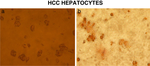
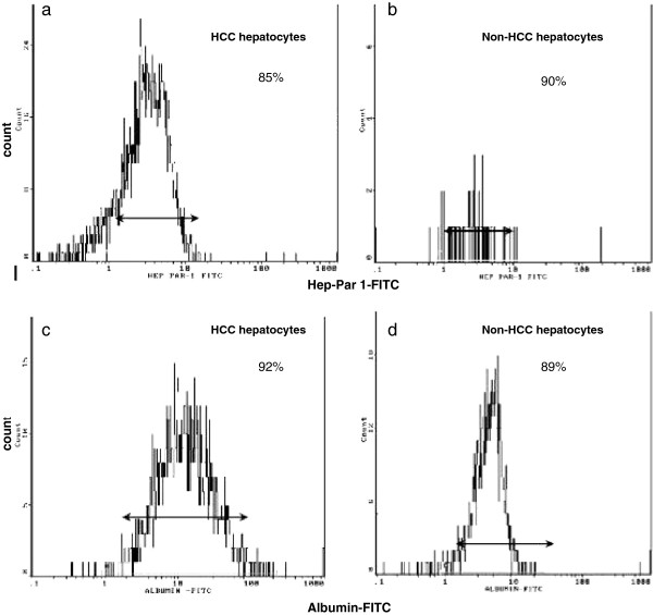
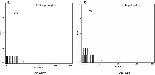
Methods: Tumor (HCC) and non-tumor (non-HCC) hepatocytes were isolated from the liver resection specimens of 11 patients operated for HCC, while PBMCs were retrieved immediately prior to surgery. Four biopsies were obtained from patients with no liver disease who had surgery for non malignant tumor (normal hepatocytes). Hepatocytes were either cultured alone (monoculture) or co-cultured with PBMCs. Flow cytometry measurements for MHC class II expression, apoptosis, necrosis and viability (7AAD) were performed 24 h, 48 h and 72 h in co-culture and monocultures.

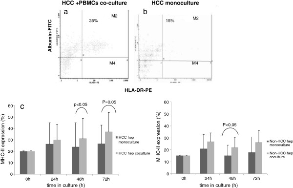
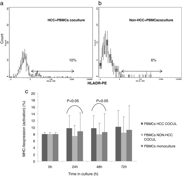
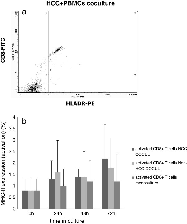
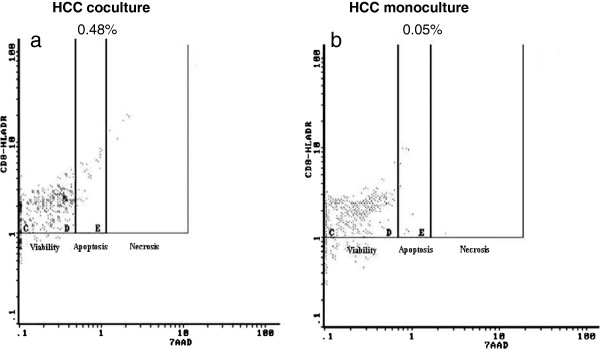
Results: HCC and non-HCC hepatocytes exhibited increased MHC-II expression at 48h and 72h in co-culture with PBMCs as compared to monoculture, with MHC II-expressing HCC hepatocytes showing increased viability at 72 h. PBMCs showed increased MHC-II expression (activation) in co-culture with HCC as compared to non-HCC hepatocytes at all time points. Moreover, CD8+ T cells had significantly increased apoptosis and necrosis at 48h in co-culture with HCC hepatocytes as compared to monocultures. Interestingly, MHC-II expression on both HCC and non-HCC hepatocytes in co-culture was positively correlated with the respective activated CD8+ T cells.

Conclusions: We have established an in vitro co-culture model to study interactions between autologous PBMCs and primary HCC and non-HCC hepatocytes. This direct interaction leads to increased antigen presenting ability of HCC hepatocytes, activation of PBMCs with a concomitant apoptosis of activated CD8+ T cells. Although, a partially effective immune response against HCC exists, still tumor hepatocytes manage to escape.


Figures

Figure 1
Figure 1
a) Representative photos of HCC hepatocytes in culture 48 hours after isolation (magnification x 20) and b) HCC hepatocytes stained with Hep Par-1 after isolation (immunocytochemistry) (magnification × 20).
Figure 2
Figure 2
Flow cytometry diagrams presenting: a) percentage of HCC-tumor hepatocytes and b) percentage of non-HCC hepatocytes stained positive for Hep Par-1, using the secondary antibody Zenon-488, after isolation c) percentage of HCC-tumor and d) percentage of non-HCC hepatocytes stained for human albumin-FITC.
Figure 3
Figure 3
After isolation tumor, non-tumor and normal hepatocytes were stained for CD3 and CD14 markers and analyzed by flow cytometry in order to verify the purity of the hepatocyte culture was performed; representative flow cytometric diagram of a) CD3-FITC and b) CD14-PE negative staining in tumor hepatocytes.
Figure 4
Figure 4
Representative flow cytometry scatterplot depicting the increased expression of MHC II on HCC hepatocytes (M region) a) 48 hours in co-culture with autologous PBMCs vs. b) 48 hours in monoculture. Representative graph showing the percentage of MHC II expression on c) HCC and d) non-HCC hepatocytes in co-culture compared to hepatocyte monocultures. Time point zero (0 h) depicts MHC II expression on hepatocytes directly after isolation.
Figure 5
Figure 5
Flow cytometric diagrams showing total PBMCs activation (MHC-II expression) after 48 h in co-culture with a) HCC and b) non-HCC hepatocytes. c) PBMCs exhibited significant activation at 24 h and 48 h when co-cultured with HCC hepatocytes compared to non-HCC hepatocytes. Also, the activation of PBMCs in HCC co-culture was higher compared to PBMCs monoculture. Time point zero (0 h) depicts activation of total PBMCs after isolation (0 h).
Figure 6
Figure 6
a) Representative flow cytometric diagram showing the increased expression of MHC class II on CD8+ T cells (activated) at 72 h HCC co-culture, b) CD8+ T cells exhibited higher activation at all time points compared to control PBMCs culture with a peak at 72 h. Also, the activation of CD8+ T cells in both HCC and non-HCC cocultures was higher compared to CD8+ T cells activation after isolation (0h).
Figure 7
Figure 7
Flow cytometric diagrams showing the % of activated CD8+ T cells (CD8+ MHC II) that are apoptotic (D region) and necrotic (E region) in a) HCC co-cultures and b) in control PBMCs cultures.

References

    1. Korangy F, Ormandy LA, Bleck JS, Klempnauer J, Wilkens L, Manns MP, Greten TF. Spontaneous tumor-specific humoral and cellular immune responses to NY-ESO-1 in hepatocellular carcinoma. Clin Cancer Res. 2004;10:4332–4341. doi: 10.1158/1078-0432.CCR-04-0181. - DOI - PubMed
    1. Mizukoshi E, Nakamoto Y, Marukawa Y, Arai K, Yamashita T, Tsuji H, Kuzushima K, Takiguchi M, Kaneko S. Cytotoxic T cell responses to human telomerase reverse transcriptase in patients with hepatocellular carcinoma. Hepatology. 2006;43:1284–1294. doi: 10.1002/hep.21203. - DOI - PubMed
    1. Liu Y, Daley S, Evdokimova VN, Zdobinski DD, Potter DM, Butterfield LH. Hierarchy of alpha fetoprotein (AFP)-specific T cell responses in subjects with AFP-positive hepatocellular cancer. J Immunol. 2006;177:712–721. - PMC - PubMed
    1. Wada Y, Nakashima O, Kutami R, Yamamoto O, Kojiro M. Clinicopathological study on hepatocellular carcinoma with lymphocytic infiltration. Hepatology. 1998;27:407–417. doi: 10.1002/hep.510270214. - DOI - PubMed
    1. Pang YL, Zhang HG, Peng JR, Pang XW, Yu S, Xing Q, Yu X, Gong L, Yin YH, Zhang Y, Chen WF. The immunosuppressive tumor microenvironment in hepatocellular carcinoma. Cancer Immunol Immunother. 2009;58:877–886. doi: 10.1007/s00262-008-0603-5. - DOI - PMC - PubMed

Publication types

MeSH terms

Substances