Normal variation of magnetic resonance T1 relaxation times in the human population at 1.5 T using ShMOLLI
- PMID: 23331520
- PMCID: PMC3610210
- DOI: 10.1186/1532-429X-15-13
Normal variation of magnetic resonance T1 relaxation times in the human population at 1.5 T using ShMOLLI
Abstract
Background: Quantitative T1-mapping is rapidly becoming a clinical tool in cardiovascular magnetic resonance (CMR) to objectively distinguish normal from diseased myocardium. The usefulness of any quantitative technique to identify disease lies in its ability to detect significant differences from an established range of normal values. We aimed to assess the variability of myocardial T1 relaxation times in the normal human population estimated with recently proposed Shortened Modified Look-Locker Inversion recovery (ShMOLLI) T1 mapping technique.
Methods: A large cohort of healthy volunteers (n = 342, 50% females, age 11-69 years) from 3 clinical centres across two countries underwent CMR at 1.5T. Each examination provided a single average myocardial ShMOLLI T1 estimate using manually drawn myocardial contours on typically 3 short axis slices (average 3.4 ± 1.4), taking care not to include any blood pool in the myocardial contours. We established the normal reference range of myocardial and blood T1 values, and assessed the effect of potential confounding factors, including artefacts, partial volume, repeated measurements, age, gender, body size, hematocrit and heart rate.
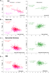
Results: Native myocardial ShMOLLI T1 was 962 ± 25 ms. We identify the partial volume as primary source of potential error in the analysis of respective T1 maps and use 1 pixel erosion to represent "midwall myocardial" T1, resulting in a 0.9% decrease to 953 ± 23 ms. Midwall myocardial ShMOLLI T1 was reproducible with an intra-individual, intra- and inter-scanner variability of ≤2%. The principle biological parameter influencing myocardial ShMOLLI T1 was the female gender, with female T1 longer by 24 ms up to the age of 45 years, after which there was no significant difference from males. After correction for age and gender dependencies, heart rate was the only other physiologic factor with a small effect on myocardial ShMOLLI T1 (6ms/10bpm). Left and right ventricular blood ShMOLLI T1 correlated strongly with each other and also with myocardial T1 with the slope of 0.1 that is justifiable by the resting partition of blood volume in myocardial tissue. Overall, the effect of all variables on myocardial ShMOLLI T1 was within 2% of relative changes from the average.
Conclusion: Native T1-mapping using ShMOLLI generates reproducible and consistent results in normal individuals within 2% of relative changes from the average, well below the effects of most acute forms of myocardial disease. The main potential confounder is the partial volume effect arising from over-inclusion of neighbouring tissue at the manual stages of image analysis. In the study of cardiac conditions such as diffuse fibrosis or small focal changes, the use of "myocardial midwall" T1, age and gender matching, and compensation for heart rate differences may all help to improve the method sensitivity in detecting subtle changes. As the accuracy of current T1 measurement methods remains to be established, this study does not claim to report an accurate measure of T1, but that ShMOLLI is a stable and reproducible method for T1-mapping.
Figures


Similar articles
-
Shortened Modified Look-Locker Inversion recovery (ShMOLLI) for clinical myocardial T1-mapping at 1.5 and 3 T within a 9 heartbeat breathhold.J Cardiovasc Magn Reson. 2010 Nov 19;12(1):69. doi: 10.1186/1532-429X-12-69. J Cardiovasc Magn Reson. 2010. PMID: 21092095 Free PMC article.
-
Systolic ShMOLLI myocardial T1-mapping for improved robustness to partial-volume effects and applications in tachyarrhythmias.J Cardiovasc Magn Reson. 2015 Aug 28;17(1):77. doi: 10.1186/s12968-015-0182-5. J Cardiovasc Magn Reson. 2015. PMID: 26315682 Free PMC article.
-
Comparison of different cardiovascular magnetic resonance sequences for native myocardial T1 mapping at 3T.J Cardiovasc Magn Reson. 2016 Oct 4;18(1):65. doi: 10.1186/s12968-016-0286-6. J Cardiovasc Magn Reson. 2016. PMID: 27716344 Free PMC article.
-
Native Myocardial T1 Mapping, Are We There Yet?Int Heart J. 2016 Jul 27;57(4):400-7. doi: 10.1536/ihj.16-169. Epub 2016 Jul 11. Int Heart J. 2016. PMID: 27396560 Review.
-
Normal values for cardiovascular magnetic resonance in adults and children.J Cardiovasc Magn Reson. 2015 Apr 18;17(1):29. doi: 10.1186/s12968-015-0111-7. J Cardiovasc Magn Reson. 2015. PMID: 25928314 Free PMC article. Review.
Cited by
-
Reference values of myocardial native T1 and T2 mapping values in normal Indian population at 1.5 Tesla scanner.Int J Cardiovasc Imaging. 2022 Nov;38(11):2403-2411. doi: 10.1007/s10554-022-02648-2. Epub 2022 Jun 27. Int J Cardiovasc Imaging. 2022. PMID: 36434341
-
Reference ranges of myocardial T1 and T2 mapping in healthy Chinese adults: a multicenter 3T cardiovascular magnetic resonance study.J Cardiovasc Magn Reson. 2023 Nov 16;25(1):64. doi: 10.1186/s12968-023-00974-5. J Cardiovasc Magn Reson. 2023. PMID: 37968645 Free PMC article.
-
Establishment and validation of an extracellular volume model without blood sampling in ST-segment elevation myocardial infarction patients.Eur Heart J Imaging Methods Pract. 2024 Jun 10;2(1):qyae053. doi: 10.1093/ehjimp/qyae053. eCollection 2024 Jan. Eur Heart J Imaging Methods Pract. 2024. PMID: 39224096 Free PMC article.
-
Qualitative and Quantitative Stress Perfusion Cardiac Magnetic Resonance in Clinical Practice: A Comprehensive Review.Diagnostics (Basel). 2023 Jan 31;13(3):524. doi: 10.3390/diagnostics13030524. Diagnostics (Basel). 2023. PMID: 36766629 Free PMC article. Review.
-
Multi-site comparison of parametric T1 and T2 mapping: healthy travelling volunteers in the Berlin research network for cardiovascular magnetic resonance (BER-CMR).J Cardiovasc Magn Reson. 2023 Aug 14;25(1):47. doi: 10.1186/s12968-023-00954-9. J Cardiovasc Magn Reson. 2023. PMID: 37574535 Free PMC article.
References
-
- Fullerton GD. In: Magnetic resonance imaging. 2. Stark DD, Bradley WG, editor. St. Louis; London: Mosby; 1992. Physiologic basis of magnetic relaxation; pp. 88–108.
-
- Bottomley PA, Foster TH, Argersinger RE, Pfeifer LM. A review of normal tissue hydrogen NMR relaxation times and relaxation mechanisms from 1–100 MHz: dependence on tissue type, NMR frequency, temperature, species, excision, and age. Med Phys. 1984;11(4):425–448. doi: 10.1118/1.595535. - DOI - PubMed
-
- Dall’Armellina E, Piechnik S, Ferreira V, Si QL, Robson M, Francis J, Cuculi F, Kharbanda R, Banning A, Choudhury R. Cardiovascular magnetic resonance by non contrast T1 mapping allows assessment of severity of injury in acute myocardial infarction. J Cardiovasc Magn Reson. 2012;14(1):15. doi: 10.1186/1532-429X-14-15. - DOI - PMC - PubMed
-
- Ferreira V, Piechnik S, Dall’Armellina E, Karamitsos T, Francis J, Choudhury R, Kardos A, Friedrich M, Robson M, Neubauer S. The diagnostic performance of non-contrast T1-mapping in patients with acute myocarditis on cardiovascular magnetic resonance imaging. J Cardiovasc Magn Reson. 2012;14(Suppl 1):P179. - PMC - PubMed
Publication types
MeSH terms
Grants and funding
LinkOut - more resources
Full Text Sources
Other Literature Sources
Research Materials

