Review
. 2013 Jan;15(1):63-79.
doi: 10.1111/ede.12015.

Evolution and development of the tetrapod auditory system: an organ of Corti-centric perspective

Affiliations
Review

Evolution and development of the tetrapod auditory system: an organ of Corti-centric perspective

Bernd Fritzsch et al. Evol Dev. 2013 Jan.

Abstract

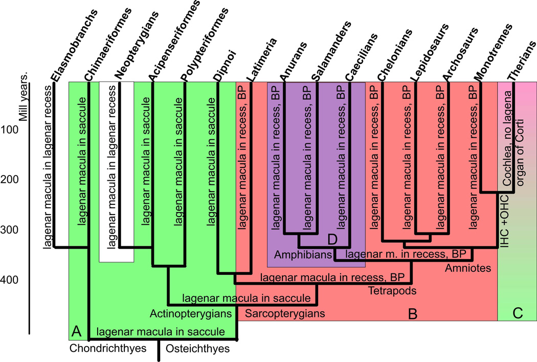
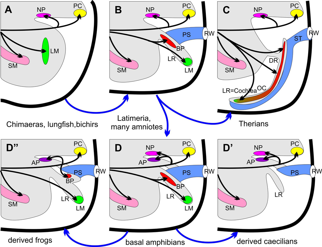
The tetrapod auditory system transmits sound through the outer and middle ear to the organ of Corti or other sound pressure receivers of the inner ear where specialized hair cells translate vibrations of the basilar membrane into electrical potential changes that are conducted by the spiral ganglion neurons to the auditory nuclei. In other systems, notably the vertebrate limb, a detailed connection between the evolutionary variations in adaptive morphology and the underlying alterations in the genetic basis of development has been partially elucidated. In this review, we attempt to correlate evolutionary and partially characterized molecular data into a cohesive perspective of the evolution of the mammalian organ of Corti out of the tetrapod basilar papilla. We propose a stepwise, molecularly partially characterized transformation of the ancestral, vestibular developmental program of the vertebrate ear. This review provides a framework to decipher both discrete steps in development and the evolution of unique functional adaptations of the auditory system. The combined analysis of evolution and development establishes a powerful cross-correlation where conclusions derived from either approach become more meaningful in a larger context which is not possible through exclusively evolution or development centered perspectives. Selection may explain the survival of the fittest auditory system, but only developmental genetics can explain the arrival of the fittest auditory system. [Modified after (Wagner 2011)].


Figures

Figure 1
Figure 1
This figure displays a consensus diagram of multiple cladistic analyses pointing out the likely relationship between different sarcopterygian and actinopterygian taxa and in different shades the major morphological changes. Note that lungfish resemble basal actinopterygians and Chondrichthyes, in particular ratfish, in great detail in that the lagenar macula is together with the saccular macula in the saccular recess (highlighted in light green, A). The derived conditions of tetrapods, apparently shared with the coelacanth Latimeria, is the possession of a lagenar recess with the lagenar macula at the tip, the formation of a basilar papilla and a perilymphatic sac to a round window in the posterior wall of the otocyst (highlighted in light red, B). Some derived Chondrichthyes and actinopterygians also have a lagenar recess with a lagenar macula in it (shown in white). All amphibians have a shared derived character, the amphibian papilla in its own recess (highlighted in light lilac, D). Salamanders and frogs have lost the neglected papilla and some salamanders and caecilians have lost the basilar papilla which may be in its own recess that comes off the lagenar recess. Some caecilians have lost the lagenar macula but retain the lagenar recess devoid of any hair cells or innervation. Ancestral mammals evolved a basilar papilla with two types of hair cells arranged in multiple rows, inner and outer hair cells (IHC, OHC). Therian mammals either lost or transformed the lagenar macula into the apex of the organ of Corti in a greatly elongated lagenar recess, now referred to as the cochlea or cochlear duct (shown in shades of light green to light red, C). Cladistic relationships compiled from (Shubin et al. 2009; Amemiya et al. 2010; Raincrow et al. 2011; Shan and Gras 2011).
Figure 2
Figure 2
This diagram displays the basic changes in the posterior part of the ear in the sarcopterygian lineage. The plesiomorphic organization or shared primitive pattern (A) is found in lungfish, basal actinopterygians (polypteriformes, holocephalens, chondrosteans, holosteans) and basal Chondrichthyes (chimaeras). The characteristic features are the presence of a nerve branch of the fibers to the posterior canal crista (PC) extending to the neglected papilla (NP), a separate nerve twig going to the distinct lagenar macula (LM) in the saccular recess posterior to the saccular macula (SM). Note that there appears to be no opening other than for nerves in the otic capsule (bold line) and no specialization of the perilymphatic space enabling pressure differences to generate relative movements between sensory epithelia and their extracellular covering (otoconia, cupula). The derived condition (B) is found in Latimeria and amniotes, excluding therian mammals. The derived condition is characterized by the formation of a lagenar recess (LR), translocation of the lagenar macula (LM) into the lagenar recess, formation of the basilar papilla (BP) at the orifice of the lagenar recess, and the formation of a perilymphatic sac (PS) that connects the basilar papilla functionally with an opening in the otocyst wall, the round window (RW). Note that the innervation of the basilar papilla is via a nerve branch coming off the lagenar macula innervation. Further derived from this basic tetrapod condition are basic amphibians (many caecilians) through the formation of a recess for the amphibian papilla (AP) and the translocation of a part of the neglected papilla into this unique recess as an amphibian papilla (D). Salamanders and frogs have all lost the neglected papilla (D”) and most salamanders and all frogs have evolved a second recess from the lagenar recess that contains the basilar papilla thus generating the derived conditions of two saccular recesses (in addition to the lagenar recess) each with its own sensory epithelium (amphibian and basilar papilla). Note also that the derived condition of innervation for the basilar papilla is via a nerve coming off the nerve to the posterior canal crista that can be reconciled through intermediates. Finally, loss of the lagenar macula occurred independently in therian mammals where the anlage might have become incorporated into the elongated basilar papilla now referred to as the organ of Corti (green tip of the red basilar papilla) due to its unique organization of hair cells (C), which is connected via narrow canal, the ductus reuniens (DR) to the saccular recess (D’). Loss of lagenar macula also occurred in some caecilians where both the basilar papilla and the lagenar macula are lost (D’) but a short lagenar recess is retained. Modified after (Lewis et al. 1985; Fritzsch and Wake 1988; Fritzsch 1992).
Figure 3
Figure 3
This figure displays the loss of neurosensory development in the cochlea duct (CD) of two mutant mouse lines, a conditional deletion of Dicer 1 with Foxg1-cre (A,B) and deletion of Gata3 (C). Note that in either case hair cells and innervation exists to vestibular epithelia interpreted as utricle (U) and saccule (S), identifiable by the endolymphatic duct (ED) emanating from it. The cochlear duct is devoid of both hair cells and innervation other than autonomic fibers. The middle panel shows a diagram of the normal innervation and organization of the organ of Corti and the cochlear duct with the scala tympani (ST) attached (D), the loss of any innervation and hair cell formation in the two mutant lines but retention of the cochlear duct (E) and the loss of the basilar papilla and lagenar macula in a caecilian (F). The right column shows 3D reconstruction of confocal images of an E16.5 wildtype ear with Amira software (G), the remaining ‘ear’ of a conditional deletion of Dicer with the cochlear duct extending from the undivided upper part (H) and the partially developed ear of a Gata3 null mouse with a cochlear duct connected via a ductus reunions to the vestibular part of the ear. For abbreviations see Fig. 2. Images taken from (Duncan et al. 2011; Kersigo et al. 2011).
Figure 4
Figure 4
The distribution of the six mammalian sensory epithelia and the neglected papilla (NP) is shown for a wildtype ear (A) and Lmx1a mutant ear (B). Note that there is continuity of utricle (U), saccule (S), and organ of Corti (OC) in the mutants. Also, the cochlea has a basal part that is converted into a more vestibular like system and an apex that is more cochlear like indicating different effects of Lmx1a along the length of the cochlea. There is a great enlargement of the posterior canal crista as well as the two patches of neglected papilla (NP) extending into the wide cochlear duct. The middle column shows the transformation of the ear in the Lmx1a null mutant. The saccular macula fuses with the organ of Corti, and there is no approximation of the scala tympani with the base of the organ of Corti but only with the apex (D). There is a dramatic increase in size of the posterior canal crista and neglected papilla. AC, anterior canal crista; HC, horizontal canal crista; DR, ductus reuniens; RW, round window; SM, scala media; UCSF; utriculo-saccular foramen. Modified after (Nichols et al. 2008)
Figure 5
Figure 5
Three dimensional reconstruction of the inner ear shows the different regions of the cochlea in wildtype (a). Deletion of N-Myc results in severe truncation of the cochlear duct and aberration in the appropriate segregation of the different regions of the cochlea (b). In particular, the apex of the N-Myc CKO cochlea forms a circular sac-like structure resembling a ‘lagena’ (yellow arrow in b’). Dye tracing from the cochlear nucleus shows afferent innervation to the abnormally formed circular sac or ‘transformed lagena’. Double knockout (dCKO) of N-Myc and L-Myc adds to the severity in the N-Myc CKO phenotype (c-c”‘). Myo7a immunohistochemistry shows abnormal fusion of the hair cells from the basal tip with the saccular hair cells (white arrows in c-c”). The dCKO also shows formation of the segregated circular sac from the apical tip of the cochlea (yellow arrows in c, c”‘). The shortened cochlea forms multiple rows of hair cells in the apex (c”). Neurofilament immunohistochemistry shows the overshooting of the fibers to this circular sac (c”‘). A, apex; B, base; CD, cochlear duct; L, lagena; OC, organ of Corti; S, saccule; SV, scala vestibuli; ST, scala tympani; U, utricle. Bar indicates 100 µm. Modified after (Kopecky et al. 2011; Pan et al. 2011; Kopecky et al. 2012).
Figure 6
Figure 6
This scheme shows the evolution of the innervation of the postero-ventral aspect of the gnathostome ear and the polarity pattern in three sensory epithelia, the saccular macula (SM), the lagenar macula (LM) and the basilar papilla (BP)/organ of Corti (OC). Note that basal sarcopterygians (lungfish) and actinopterygians (bichir) show a detailed identity in the lagenar hair cell polarity organization (A) that is distinct from that of tetrapods (B, B’) and also from the different pattern in many derived bonyfish that have a lagenar macula in its own recess (A’). Therian mammals (C) differ from monotremes (B) by the possible fusion of the lagenar macula with the basilar papilla (green and red) forming the organ of Corti. Note that innervation is highly stereotyped and similar across vertebrates except for amphibians with the innervation of the amphibian papilla of a neglected papilla (NP) twig (if the later exist) and formation of a new posterior twig to the basilar papilla coming off the posterior canal nerve in derived frogs (B’). A similar pattern of innervation can be generated in mice using a knockin of the neurotrophin Bdnf into the Ntf3 locus, turning the basal turn of the organ of Corti innervation into an innervation resembling the derived frog basilar papilla.

References

    1. Abu-Daya A, Khokha MK, Zimmerman LB. The hitchhiker's guide to Xenopus genetics. Genesis. 2012;50(3):164–175. - PMC - PubMed
    1. Ahmed M, Wong Elaine ÂYM, Sun J, Xu J, Wang F, Xu P-X. Eya1-Six1 Interaction Is Sufficient to Induce Hair Cell Fate in the Cochlea by Activating Atoh1 Expression in Cooperation with Sox2. Developmental Cell. 2012;22(2):377–390. - PMC - PubMed
    1. Ahn KJ, Passero F, Jr, Crenshaw EB., 3rd Otic mesenchyme expression of Cre recombinase directed by the inner ear enhancer of the Brn4/Pou3f4 gene. Genesis. 2009;47(3):137–141. - PubMed
    1. Amemiya CT, Powers TP, Prohaska SJ, Grimwood J, Schmutz J, Dickson M, Miyake T, Schoenborn MA, Myers RM, Ruddle FH, Stadler PF. Complete HOX cluster characterization of the coelacanth provides further evidence for slow evolution of its genome. Proc Natl Acad Sci U S A. 2010;107(8):3622–3627. - PMC - PubMed
    1. Basch ML, Ohyama T, Segil N, Groves AK. Canonical Notch signaling is not necessary for prosensory induction in the mouse cochlea: insights from a conditional mutant of RBPjkappa. J Neurosci. 2011;31(22):8046–8058. - PMC - PubMed

Publication types

LinkOut - more resources