. 2013 May;37(5):503-9.
doi: 10.1016/j.leukres.2012.12.013. Epub 2013 Jan 17.

Adipose tissue attracts and protects acute lymphoblastic leukemia cells from chemotherapy

Affiliations

Adipose tissue attracts and protects acute lymphoblastic leukemia cells from chemotherapy

Rocky Pramanik et al. Leuk Res. 2013 May.

Abstract

Obesity is associated with an increased risk of acute lymphoblastic leukemia (ALL) relapse. Using mouse and cell co-culture models, we investigated whether adipose tissue attracts ALL to a protective microenvironment. Syngeneically implanted ALL cells migrated into adipose tissue within ten days. In vitro, murine ALL cells migrated towards adipose tissue explants and 3T3-L1 adipocytes. Human and mouse ALL cells migrated toward adipocyte conditioned media, which was mediated by SDF-1α. In addition, adipose tissue explants protected ALL cells against daunorubicin and vincristine. Our findings suggest that ALL migration into adipose tissue could contribute to drug resistance and potentially relapse.


Conflict of interest statement

Conflict of Interest:

The authors have no conflicts of interest to disclose.

Figures

Figure 1
Figure 1
Migration of ALL cells toward adipose tissue. Summary of flow cytometry analysis of ALL cells (GFP positive) in blood, bone marrow, various tissues and SVF of adipose tissue. Six obese (HF) and six control (LC) mice were transplanted. (A) The number of mice that had detectable amount of ALL cells is shown. The detection limit is set for each tissue using samples from a non-transplanted mouse. (B) Quantification of ALL cells in the sites detected in (A) analyzed by flow cytometry. Percentages are calculated by dividing the amount of GFP+ cells by the total number of live cells in mice with detectable amounts of ALL cells. No ALL cells were detected in muscle (N.D.). (*p<0.05, HF visceral vs. LC visceral) (C) Weight of fat depots from obese and lean mice. (***p<0.001) (D) Tissue explants from obese mice were cultured for 2 days and 8093 ALL cells were plated in transwells above the explants and incubated for 90 minutes. % migration is calculated as number of cells in bottom chamber divided by total number of cells counted in both chambers. (*p<0.05 and **p<0.01 vs. no tissue, n=3)
Figure 2
Figure 2
Migration of ALL cells to adipocytes and ACM. (A) Migration of 8093 murine ALL cells toward pre-adipocyte conditioned medium (FCM), ACM, pre-adipocyte feeder layer (Fibro), and adipocyte feeder layer (Adipo) of 3T3-L1 cells and OP9 cells. Percentage of migrated cells is calculated as before. Control condition was performed with RPMI medium in both chambers. (*p<0.05 and **p<0.01 vs. control, n=3) (B) ACM causes both directional and non-directional movement of 8093 ALL cells. Top and bottom chambers are filled with either RPMI (C) or ACM (A). (**p<0.01 and *p<0.05 vs. medium, n=4) (C) Time course of 8093 ALL migration toward ACM (n=2).
Figure 3
Figure 3
Migration of ALL cells toward SDF-1α and blocking by AMD3100. (A) Human ALL cell lines were incubated in control medium, and migration toward control, ACM, or SDF-1α (60 ng/mL) was quantified. (B) Migration of primary human ALL cell strains migrate toward 60 ng/mL SDF1α. (*p<0.05, **p<0.01, and ***p<0.001 vs. control, n=3) (C) ELISA quantification of SDF-1α secretion by 3T3-L1 and OP9 pre-adipocytes and adipocytes, measured in media conditioned for 48 hours. (**p<0.01, n=3) (D) Inhibition of migration of 8093 cells towards SDF-1α (n=3) or ACM (n=4) after pre-incubation of cells with the indicated amounts of AMD3100. (*p<0.05, **p<0.01, and ***p<0.001 vs. zero dose of AMD3100) (E) Migration of 8093 cells towards OP9 FCM and ACM (n=3) after pre-incubation of cells with the indicated amounts of AMD3100 (**p<0.01, and ***p<0.001 vs. zero dose of AMD3100). (F) Western blot for CXCR4 in leukemia cell lines. β-actin is included as loading control.
Figure 4
Figure 4
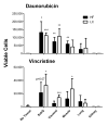
Protection of ALL cells by fat explants against daunorubicin and vincristine. Weight-matched tissue explants (subcutaneous fat, visceral fat, muscle, lungs, and kidneys) from control and obese mice (n=6 each) were excised after cardiac perfusion. The doses of daunorubicin (19nM) and vincristine (3nM) were EC99 doses in the control conditions without tissue explants. ALL cells were quantified using trypan blue exclusion after 48 hours of drug treatment (*p<0.05, **p<0.01, and * **p<0.001 vs. no tissue control).

References

    1. Pui CH, Carroll WL, Meshinchi S, Arceci RJ. Biology, risk stratification, and therapy of pediatric acute leukemias: an update. J Clin Oncol. 2011;29:551–565. - PMC - PubMed
    1. Fielding A. The treatment of adults with acute lymphoblastic leukemia. Hematology Am Soc Hematol Educ Program. 2008:381–389. - PubMed
    1. Buchanan GR. Diagnosis and management of relapse in acute lymphoblastic leukemia. Hematol Oncol Clin North Am. 1990;4:971–995. - PubMed
    1. Kurtova AV, Balakrishnan K, Chen R, Ding W, Schnabl S, Quiroga MP, et al. Diverse marrow stromal cells protect CLL cells from spontaneous and drug-induced apoptosis: development of a reliable and reproducible system to assess stromal cell adhesion-mediated drug resistance. Blood. 2009;114:4441–4450. - PMC - PubMed
    1. Mudry RE, Fortney JE, York T, Hall BM, Gibson LF. Stromal cells regulate survival of B-lineage leukemic cells during chemotherapy. Blood. 2000;96:1926–1932. - PubMed

Publication types

MeSH terms

LinkOut - more resources